
Page 229 - 2019_03-Haematologica-web
P. 229
Somatic mosaicism at different stages of ontogenesis
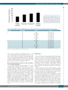
Figure 2. Red cell C antigen (RH2) expression levels of proposita A and B compared to normal controls. Mean fluorescence intensities of erythrocytes indi- rectly stained with polyclonal anti-C after subtraction of negative control obtained with ccddee cells are shown. CcDdee (n=3) and CcDdee (n=3) control val- ues are depicted as average with standard deviation.
Table 2. Molecular RHCE*c analysis and RHd typing of single erythroid progenitor cells of both individuals with Rh phenotype splitting.
Proposita BFU-E colony
A A1 A2 A3 A4 A5 A6
B B1 B2 B3 B4 B5 B6
LOH: loss of heterozygosity; BFU-E: erythropoietic burst-forming units.
These results underlined the haplotypic nature of the observed blood group anomaly and indicated the co-exis- tence of 2 RBC lines: 1 of normal c-positive phenotype encoded by 2 parental RH haplotypes (CDe/cde) and a sec- ond with c-negative phenotype encoded by the LOH- modified RHCE*c-negative parental haplotype only (homozygous CDe/CDe or hemizygous CDe/---).
Rh blood group anomaly caused by somatic recombina- tion-associated duplication
To determine whether the Rhc-negative cell clone resulted from a hemizygous deletion (CDe/---) or a more complex somatic recombination-associated duplication of the CDe haplotype, dual-color FISH analyses on fixed peripheral blood cells obtained from both studied individ- uals were performed. In both proposita A and B, FISH analysis showed the diploid presence of the RH loci in all segmented and round nuclei (Figure 4). Despite there being no proof by chromosomal sequencing, these results indicated somatic recombination-associated loss of the RHCE*c-positive/RHD-negative and duplication of the RHCE*c-negative/RHD-positive haplotype as cause for the observed RBC phenotype splitting. Hence, the LOH- affected RHCE*c-negative cell lines of both propositae most probably harbored homozygous CDe/CDe haplo- types.
RHCE*c
LOH heterozygous heterozygous LOH heterozygous LOH heterozygous LOH LOH heterozygous heterozygous LOH
Discussion
RHd
absent (DD or D-) heterozygous Dd heterozygous Dd absent (DD or D-) heterozygous Dd absent (DD or D-) heterozygous Dd absent (DD or D-) absent (DD or D-) heterozygous Dd heterozygous Dd absent (DD or D-)
Two individuals with an unexplained mixed-field agglu- tination in routine serological Rhc typing have been observed. Common causes of mixed Rhc phenotype, such as RBC transfusion or hematopoietic stem cell transplan- tation, were ruled out in the 2 propositae. Extended molecular testing was performed to define the underlying mechanism of this condition. Microsatellite analysis across different chromosomes excluded spontaneous chimerism known to bring about mixed blood group phe- notypes.27
Using chromosome 1 microsatellite markers, somatic mosaicism with partial haploid loss of 1p involving the RH locus was found to be responsible for the observed Rhc phenotype anomaly in both individuals studied, encom- passing at least 26.7 and 42.4 Mb, respectively.
The high red cell expression of the antithetical C (RH2) antigen, nearly approaching levels of RHCE*C homozy- gous controls, indicates that the deletion of a part of 1p (eliminating the RHCE*C allele) has been repaired by a duplication of homologous stretches of the other chromo- some harboring the RHCE*C allele. This view is further supported by the FISH results, showing the uniform pres- ence of two RH loci in all examined cell nuclei. Accordingly, also LOH-affected cells did not show an RH
haematologica | 2019; 104(3)
635
