
Page 228 - 2019_03-Haematologica-web
P. 228
E.M. Dauber et al.
Table 1. Blood group phenotypes of the 2 propositae with spontaneous c antigen (RH4) mixed-field typing.
Proposita
A
B
Rh
D+C+E-e+Cw-
D+C+E-e+Cw-
ABO MNS
A M-N+S-s+
A M+N+S+s+
Blood group phenotype P1Pk Lutheran
c± (53% c+)
c± (50% c+)
P1- Lu(a-b+)
P1+ Lu(a-b+)
Kell
K-k+, Kp(a-b+)
K+k+, Kp(a-b+)
Duffy
Fy(a+b+)
Fy(a+b+)
Kidd
Jk(a+b+)
Jk(a+b-)
±: mixed-field agglutination.
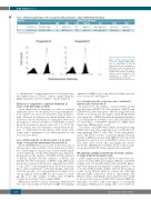
Figure 1. Flow cytometric analysis of red cell c antigen (RH4) expres- sion of propositae A and B. Immunofluorescence histograms of erythrocytes indirectly stained with polyclonal anti-c are shown. Note c- negative and c-positive cell subpop- ulations (black histograms). For comparison, a CcDdee control (open histograms) is included.
1). Antithetical C antigen expression of both propositae was higher than in CcDdee controls, approaching the higher quantities seen in CCDDee controls (Figure 2).
Exclusion of congenital or acquired chimerism as cause of Rh phenotype anomaly
Twin chimerism or dispermy, as well as artificial chimerism (due to blood transfusion or organ transplanta- tion) could be the reason for mixed blood group pheno- types. However, both propositae denied having a twin or a history of blood transfusions or organ grafts. Moreover, the analysis of 15 microsatellite loci with DNA of whole blood (loci located on chromosomes 2-5, 7, 8, 11-13, 16, 18, 19, and 21) ruled out chimerism: exclusively homozy- gous or well-balanced heterozygous allelic peaks were found, with a maximum of two alleles present at each locus (data not shown).
Loss of heterozygosity on chromosome 1 at an early stage of ontogenetic development in proposita A
As the RHD/RHCE loci are located on the short arm of chromosome 1, the possibility of mosaicism was tested by use of heterozygous chromosome 1 microsatellite mark- ers (for full details, see the Online Supplementary Appendix). In proposita A, the analysis of D1S468 (21 Mb telomeric of RH*D), D1S234 (0.5 Mb telomeric of RH*D), and D1S233 (5.7 Mb centromeric of RH*D) using DNA from whole blood, and sorted leukocyte subpopulations (CD4+ T cells, CD8+ T cells and granulocytes) showed in all sam- ples a clear-cut imbalance of the peak heights. This indi- cated the presence of 2 cell populations in which 1 lost one 1p segment. Such an LOH was also seen in 2 of 6 sin- gle hair roots. The analysis of DNA from 19 BFU-E colonies showed that 9 had complete LOH.
Other microsatellite loci more centromeric than D1S233 were also tested, without evidence for LOH. The minimal
expansion of LOH on 1p of the affected cell lines amount- ed to at least 26.7 Mb (Figure 3).
Loss of heterozygosity on chromosome 1 confined to myeloid cells in proposita B
In proposita B, the analysis of microsatellites in the region between D1S507 (10.3 Mb telomeric of RH*D) and D1S2890 (32.1 Mb centromeric of RH*D) using DNA from whole blood showed in all samples a peak height imbal- ance diagnostic of LOH, thus demonstrating the existence of 2 cell populations in which 1 lost 1 allele. D1S252 locat- ed centromeric of D1S2890 exhibited no LOH. Hairs showed no LOH in all loci tested.
The alleles of D1S2890 were further investigated using DNA from buccal swab, single hair roots, sorted leukocyte subpopulations (CD4+ T cells, CD8+ T cells, and granulo- cytes), and BFU-E colonies. A myeloid lineage-restricted pattern of LOH was found, with LOH detected in sorted granulocytes and in 4 out of 22 BFU-E colonies. In con- trast, hairs (n=3), buccal cells, and lymphocyte subsets showed no LOH (Figure 3). Further details of these analy- ses are provided in the Online Supplementary Appendix.
RH genotype splitting confirmed by molecular analysis of single erythroid progenitor cells
DNA samples from separate BFU-E colonies were sub- jected to real-time PCR genotyping for RHCE*c. Six BFU-E samples each of both individuals with mixed Rhc pheno- type were analyzed and displayed a similar pattern: 3 out of 6 tested BFU-E DNA samples showed heterozygous results for the RHCE*c allele; in contrast, the other half indicated LOH at this locus (Table 2). Importantly, only BFU-E colonies with RHCE*c heterozygosity were found to be also heterozygous for RHD (Dd), whereas LOH was uniformly associated with homozygous or potentially hemizygous RHD-positive typing (DD or D-) (Table 2).
634
haematologica | 2019; 104(3)
