
Page 115 - 2019_03-Haematologica-web
P. 115
TP53 subclones in AML
AB
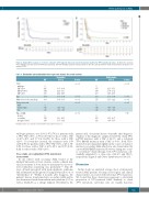
Figure 4. Kaplan-Meier analysis of event-free survival in 1537 patients with acute myeloid leukemia stratified by TP53 mutational status. (A) Event-free survival: TP53 wild-type patients versus TP53-mutated patients. (B) TP53 wild-type patients versus patients in the three groups with the defined variant allele frequencies of mutated TP53.
Table 3. Univariable and multivariable Cox regression analysis for overall survival. Univariable
Multivariable 95% CI
2.11 - 6.51 1.04 - 3.02 1.47 - 2.79
1.03 - 1.04 1.11 - 1.22
0.41 - 0.58
0.20 - 0.36
1.13 - 2.07
0.88 - 1.66
TP53
wild-type 1 VAF <20% 4.95 VAF 20%-40% 3.44 VAF >40% 3.73
Age 1.04
White blood cell count (log) 1.08
3.05 - 8.04 2.16 - 5.50 2.84 - 4.88
1.03 - 1.04 1.04 - 1.13
<0.001 <0.001 <0.001
HR 95% CI
P-value <0.001
<0.001
HR
1 3.71 1.78 2.03
1.03 1.17
P-value <0.001
<0.001 <0.001 <0.001
0.019
Cytogenetic risk
high1 1
intermediate 0.45
low 0.20 Type of AML
de novo 1 secondary 1.74 therapy-related 1.43
0.38 - 0.52
0.15 - 0.26
1.30 - 2.33
1.07 - 1.92
0.49
0.27
1 1.53 1.21
HR:hazard ratio; CI: confidence interval;VAF: variant allele frequency.
wild-type patients, was 2.03 (1.47-2.79) for patients with a TP53 VAF >40%, 1.78 (1.04-3.02) for those with a VAF of 20%-40% and 3.71 (2.11-6.51) for those with a VAF <20%. For event-free survival, the estimates were 1.89 (1.38-2.59) for patients with a TP53 VAF >40%, 2.27 (1.35- 3.80) for those with a VAF of 20%-40% and 3.57 (2.04- 6.26) for those with a VAF <20%.
Case study on longitudinal TP53 mutational assessment
In one subject with secondary AML treated at the Medical University of Graz, Austria, outside a clinical trial, serial assessment of bone marrow specimens by error-cor- rected ultradeep sequencing was performed.22,23 This 68- year old male presented with myelodysplastic syndrome and an International Prognostic Scoring System risk score of “intermediate 2”. Within 2 months after diagnosis, the patient’s disease transformed into secondary AML and he was then treated with daunorubicin and cytarabine (3+7) as well as Ida-FLAG as a salvage regimen. Nevertheless, the
patient died of resistant disease 4 months after diagnosis. Analysis of the diagnostic sample revealed the clonal TP53 p.273H mutation with a VAF of 42.3% and a subclonal TP53 Q104X mutation with a VAF of 0.4%. Both TP53- mutated clones expanded slightly in the course of transfor- mation to secondary AML which was also characterized by a novel KRAS Q61H mutation. However, during two cours- es of intensive chemotherapy, both TP53 mutations sub- stantially expanded to a clone size of 64.2% and 18.6%, respectively. (Figure 5 and Online Supplementary Table S5).
Discussion
In this study, we analyzed a large cohort of intensively treated AML patients focusing on biological and clinical characteristics associated with subclonal TP53 mutations. We found that these aberrations represent a substantial proportion of TP53-mutated AML. Similarly to clonal TP53 mutations, subclonal ones are mainly missense
haematologica | 2019; 104(3)
521
