
Page 116 - 2019_03-Haematologica-web
P. 116
K.T. Prochazka et al.
mutations and are associated with an adverse outcome as evidenced by significantly inferior complete remission, overall survival and event-free survival rates.
Results similar to those presented here have been obtained for patients with chronic lymphocytic leukemia in whom those with TP53-mutated subclones with a median VAF as low as 2.1% showed comparable clinical phenotypes and survival as poor as those with clonal mutations.24,25 Interestingly, divergent data have been pub- lished for patients with myelodysplastic syndromes.18,19 Analyzing two independent cohorts of 219 and 150 patients, also including a few cases with secondary AML, Sallman et al. stratified TP53 VAF into the same categories as defined here (>40%, 20%-40%, <20%). Importantly, there was a significant difference in overall survival between patients exhibiting TP53 mutant VAF of >40% and <20%. Whereas myelodysplastic syndrome patients with a TP53 VAF >40% had a median overall survival of 124 days, overall survival was not reached in patients with a VAF <20%. In contrast, Goel et al. investigated two cohorts of 81 and 1,117 patients and did not find any sig- nificant difference with respect to TP53 VAF and their neg- ative prognostic impact on overall survival. It was argued that one of the reasons for these discrepancies might have been the small number of TP53-mutated patients investi- gated. In addition, early therapeutic intervention leading to clonal expansion of a drug-resistant clone may also be relevant to these differences in survival.26 In our sufficient- ly powered study of 1,537 patients predominantly with de novo AML, 98 had TP53-mutated clones of various sizes. We provide evidence here that small TP53-mutated sub- clones share biological characteristics with clonal ones, such as mutation type, location of the mutation within TP53 domains and cooperating mutations. We also demonstrate that even TP53-mutated subclones, defined by a VAF <20%, have a statistically significant negative prognostic impact with respect to complete remission rate, overall survival and event-free survival. These find- ings may have implications for TP53 screening methods and future risk stratification in AML as classical Sanger sequencing with a detection limit of mutant clones set at 20% VAF should be replaced by high sensitivity next-gen- eration sequencing approaches. Furthermore, when con- firmed by others, subclonal TP53 mutations should be incorporated into future risk classification for AML.
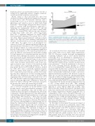
The mechanisms by which mutant p53 mediates resist- ance to cytotoxic treatments are poorly understood and possibly involve novel gain-of-function properties. TP53 mutations affect preleukemic stem cells in AML and it could be shown in vitro and in vivo that genotoxic stress leads to expansion of murine hematopoietic stem and progenitor cells exhibiting either p53 haploinsufficiency or expressing a heterozygous TP53 mutation.27-29 These data are in line with the AML case presented here, in which substantial expansion of both a clonal and a subclonal TP53 mutation was observed following high-dose chemotherapy but not during the transition from myelodysplastic syndrome to frank leukemia. Similar clinical data have recently been published for patients with lymphomas showing clonal hematopoiesis including TP53 mutations at diagnosis or before undergoing autologous hematopoietic stem cell transplantation. These individuals were at increased risk of clonal expansion and, ultimately, development of therapy- related myeloid neoplasms following intensive, lymphoma- specific chemo- and radiotherapies.30-33
Figure 5. Longitudinal mutational analyses of a patient with secondary acute myeloid leukemia showing a clonal (R273H) and a subclonal (Q104X) TP53 mutation. VAF: variant allele frequency; MDS, myelodysplastic syndrome: sAML: secondary acute myeloid leukemia; PD, progressive diesease. Bottom line: coop- erating mutations.
In our study we were able to demonstrate TP53-mutated clones with a VAF as low as 4.66% (Online Supplementary Table S1). However, recently developed next-generation sequencing techniques such as error-corrected ultradeep sequencing allow an unambiguous identificantion of mutant alleles with a frequency as low as 0.1%.34 In chronic lymphocytic leukemia, it has been demonstrated that even very small TP53-mutated clones constitute an adverse prog- nostic parameter.24,25 Given the mechanism of selection of such clones following genotoxic therapies as outlined above, it is very likely that a similar effect may also be oper- ational in AML. Another issue is related to the assessment of VAF using clinical specimens. Specification of VAF refers either to mononuclear cell fractions or with a correction to represent exclusively neoplastic cells. However, it must be taken into account that mutations affecting preleukemic stem cells in AML are being propagated into mature blood cells.35 By analyzing highly purified T-lymphocytes of diag- nostic AML specimens with TP53 mutations, we were able to demonstrate the specific aberration being present in up to 20% of these mature blood cells.27 Thus, analyzing mononuclear cell fractions may be the more comprehensive approach to assess VAF in AML and, possibly, other myeloid disorders. Finally, the diagnostic specimens in this study were derived from both bone marrow and peripheral blood (Online Supplementary Methods) raising the issue of comparability of these sources. Recently, Duncavage et al. demonstrated that the mutational landscape is conserved in peripheral blood of AML and myelodysplastic syndrome patients at diagnosis as well as during treatment.36 Similar data were obtained for TP53 mutations in patients with myelodysplastic syndromes.37
In this study we investigated the prognostic impact of subclonal TP53 mutations in intensively treated AML patients. Results may be somewhat different when assess- ing AML patients with TP53 mutations treated with hypomethylating agents such as decitabine. Although a substantial impact on overall survival could not be shown with that approach, similar response rates as those of AML patients with TP53 wild-type have been reported recently.38 It might, therefore, be necessary to prospective-
522
haematologica | 2019; 104(3)
