
Page 254 - Haematologica Vol. 110 - January 2025
P. 254
LETTER TO THE EDITOR
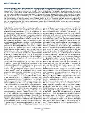
Figure 1. MAGE-A1 expression in multiple myeloma patient samples is associated with extramedullary disease and is a risk factor for overall survival. (A) Samples were classified according to the fraction of MAGE-A1-positive (MAGE-A1+) multiple myeloma (MM) cells (negative; low =1-29%; medium =30-80%; high =>80%). Proportion of the different categories of MAGE-A1 expression shown for all samples, newly diagnosed MM (NDMM) and relapsed/refractory MM (RRMM). (B) Representative immunohistochemistry staining of MM bone marrow samples with monoclonal antibody MA454 and anti-CD138 of a patient with NDMM. BM is infiltrated with 30% of MM cells of which overall 10-20% are MAGE-A1+. (C) Proportion of MAGE-A1+ MM cells categorized according to the patients cytoge- netics results. Mean and standard error of the mean shown. Kruskal-Wallis test. (D) Proportion of MAGE-A1+ MM cells depending on sample type in MAGE-A1+ samples (at least 30% positive MM cells). (E) Proportion of MAGE-A1+ MM cells depending on the presence of extramedullary disease at any time of disease course in MAGE-A1+ bone marrow samples. (F) Kaplan-Meier curves of patients with MAGE-A1 expression (at least 30% positive myeloma cells) and without (<30% positive myeloma cells) at time point of NDMM. HR: hazard ratio; CI: confidence interval.
006). T1367 expresses Vβ3, which was used as marker for T1367-transduced cells. The transduction rates, estimated by flow cytometry detection of Vβ3+/CD8+ cells 4 days af- ter transduction, were 18.6% (001), 32.7% (004) and 31.6% (006) of all CD45+/7-ADD- cells, and were stable throughout the freezing and thawing process. Only the product from patient 006 experienced a 10% decrease (Figure 2B). For patients 004 and 006 a sufficient cell proliferation after transduction was observed, reaching proliferation rates of 57-fold and 13-fold on day 12 (Figure 2C). In contrast, product 001 showed a proliferation rate of only 2-fold on day 12 (Figure 2C). We observed a strong correlation be- tween the proliferation rate from day 2 to day 5 and the transduction rate in the final product (R2=0.9441; P=0.0012; Figure 2D). The cell viability after thawing, determined by trypan blue staining, was 76.0% (001), 97.5% (004) and 93.5% (006). Due to the proliferation and viability data, the TCR-1367 T cells (patient 001) were not considered for therapy.
To evaluate safety and efficacy of TCR-1367 T cells, we conducted a one-armed, single-center, open-label, phase I clinical trial (EudraCT: 2017-001208-30). The summarized study design is shown in Figure 3A. The main inclusion criteria were age ≥18 years, relapsed and/or refractory disease requiring therapy, at least three prior lines of ther- apy, HLA-A*02:01 genotype, and at least 30% of MAGE-A1+ MM cells assessed by IHC. The primary objective was to evaluate the safety and tolerability of TCR-1367 T cells. It was planned to enroll 12 patients in four cohorts with ascending doses of TCR-1367 T cells (105; 106; 107 and 5x10⁷ cells/kg body weight [BW] ± 20%). However, based on lim- ited recruitment potential upon availability of BCMA-CAR T cells, and competing clinical studies investigating BiTE, the trial was closed by the sponsor after treating two pa- tients (patients 004 and 006). All patients provided written informed consent and the trial was approved by the local ethical committee in Berlin, Germany (17/0259-EK13). The study was conducted in accordance with principles of good clinical practice and the Declaration of Helsinki. The patient characteristics are shown in Online Supplementary Table S2. Both patients were treated in the first dosing cohort and were eligible for safety and efficacy analysis. The time between apheresis to application of TCR-1367 T cells was 64 days (004) and 55 days (006). Altogether, we
observed 18 treatment-emergent adverse events (TEAE) in the two patients with 11 classified as possibly study treat- ment-related. Four of the TEAE were CTCAE (version 4.03)13 grade 3 or 4 and two were serious AE (febrile neutropenia and cancer pain), both likely related to chemotherapy and disease progression, respectively. All AE are listed in Online Supplementary Table S3. The best response according to the IMWG criteria14 was minimal response for patient 004, while patient 006 experienced progressive disease. The time to next treatment was 110 days for patient 004 and 64 days for patient 006. In patient 004, the proportion of MAGE-A1+ MM cells subsequently decreased from 80% to 60% to 30% 3 months after administration of TCR-1367 T cells (Figure 3B). MM cell infiltration decreased from 60% to 40%. However, the fraction of MAGE-A1+ MM cells started to rise again 7 months after the administration of TCR-1367 T cells, reaching 40% (Figure 3B). As shown in Figure 3C for patient 006, no measurable effect of the TCR-1367 therapy on the MAGE-A1 expression was found. Patient 004 achieved a complete response under fol- lowing BiTE treatment and is still alive 35 months after administration of TCR-1367 T cells. Patient 006 further progressed and died 3 months after receiving the study treatment from myeloma progression and disease-related pancytopenia with an infection of unknown focus, consid- ered not related to the study treatment, resulting in an OS of 91 days. No TCR-1367 T cells could be detected by flow cytometry or quantitative polymerase chain reaction (qPCR) in pharmacokinetics samples, possibly due to the low cell number administered.
MAGE-A1, identified as a frequently expressed antigen in MM in our diagnostic study, could emerge as a valuable new target, especially considering loss of commonly targeted antigens like BCMA and GPRC5D under current therapies.6 MAGE-A1 expression was associated with EMD and lower OS, aligning with findings in other MM patient cohorts.3,15 In the phase I clinical trial investigating MAGE-A1-direct- ed TCR-1367 T cells, we treated only two patients due to premature closure of the trial by the sponsor, making it impossible to provide conclusive safety and efficacy data. While we observed no severe TEAE, durable responses were not achieved, possibly due to the low dose of TCR- 1367 T cells administered in the first dose cohort (1x10⁵ cells/kg BW).
Haematologica | 110 January 2025
246
