
Page 58 - Haematologica-5
P. 58
A. Khaibullina et al.
Methods
Mice and RONi injections
The animal protocol was approved by the Institutional Animal Care and Use Committee at the Children's National Health
System. Townes mice, here referred to as SCD mice, were obtained from the Jackson Laboratory.
Two-month old mice were injected subcutaneously with 10 mg/kg of body weight of RON inhibitor (RONi, BSM-777607, SantaCruzScientific)in2%DMSOdailyfor14consecutivedays.
A
B
C
D
E
F
P=4x10⁻⁵
P=3x10⁻⁹
790
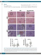
Figure 3. Glomerular macrophages infiltration and MSP1 accumulation is increased in sickle cell disease (SCD) mice. (A-D) Representative pictures of macrophages (F4/80) (A and B, red) and macrophage stimulating protein 1 (MSP1) (C and D, brown) immunostaining of renal sections. Squares show enlarged areas immunostaining (B and D). Non-specific primary antibodies (Abs) were used as a negative control. Bar sizes on the microphotographs are 100 μm (A and C) and 40 μm. (B and D). (E and F) Quantification of F4/80 positive macrophages per glomeruli cross section (E) and MSP1 accumulation in the glomeruli (F) is per- formed using ImageJ Fiji version software. Five mice per group were used for each staining. For quantification graphs, means are shown. Each dot represents a value obtained from one glomerulus cross-section. Ctrl: control.
haematologica | 2018; 103(5)
