
Page 87 - Haematologica-April 2018
P. 87
Correlation of mutations with morphological findings in AML
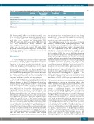
Table 2. Final transplant-adjusted multivariable model including mutational and morphological features
Stem cell transplant
Coefficient
-0.348
0.764 0.799 -0.696 0.756
Exp ( coefficient) Hazard Ratio
0.706
2.146 2.223 0.499 2.13
SE coefficient
0.271
0.319 0.248 0.248 0.447
z value
-1.283
2.391 3.22 -2.81 1.69
P
0.199
0.017 0.001 0.005 0.091
Micromegakaryocytes (score ≥3) Subclone present
NPM1 mutation
NF1 mutation
EXP: expansion.
EFS between AML-MRC versus all the other AML cases (P=0.941), nor was there any significant difference in EFS between AML-MRC versus AML-RUNX1 (P=0.253), AML-MRC versus combined AML-NPM1/AML-CEBPA (P=0.407), and AML-MRC versus AML-NOS (P=0.704). The final multivariable model indicated that micromegakaryocytes (score ≥3) and presence of a sub- clone were associated with shorter EFS while NPM1 mutation was associated with longer EFS; mutation in the RAS pathway gene NF1 was marginally associated with shorter EFS (Table 2).
Discussion
To our knowledge, this is the first study to explore the relationship of specific dysplastic findings with mutation patterns in de novo AML. We found that among the broad spectrum of dysplastic morphologies evaluated in our study, only megakaryocyte morphology showed signifi- cant associations with mutation pattern or outcome. Interestingly, we found that one specific morphological feature, megakaryocytes with separated lobes, was corre- lated with cohesin pathway and NPM1 mutations but did not impact outcome, while another morphological fea- ture, micromegakaryocytes, was associated with poor outcome, but did not correlate with any mutations.
The biological significance of morphological dysplasia in de novo AML is controversial, despite the fact that mul- tilineage dysplasia (at least 50% dysplastic cells in at least 2 hematopoietic lineages) defines a subset of cases of AML with myelodysplasia changes that lack a defining cytoge- netic abnormality or antecedent MDS.4 In our cohort of de novo AML cases, we found that cohesin pathway (and specifically STAG2) mutations were associated with greater overall megakaryocytic dysplasia and the specific finding of megakaryocytes with separated nuclear lobes. Mutations in cohesin pathway genes were present in 11% of all patients in our cohort; a higher rate than that report- ed by Thol et al. (5.9%) or in The Cancer Genome Atlas (2.5-3.5%). However, this rate might be influenced by the fact that our study included only AML cases without a his- tory of a prior myeloid neoplasm and without MDS-asso- ciated cytogenetic abnormalities.2,17 Interestingly, Thol et al. and The Cancer Genome Atlas research network found a strong association between NPM1 mutations and muta- tions in cohesin genes,2,17 and in our study 6 (31%) of 19 cases with cohesin mutations also had NPM1 mutations, and the latter were also associated with frequent megakaryocytes with separated nuclear lobes. RAS path-
way mutations were frequently seen in our cohort, being present in 58% of the cases and, similar to cohesin path- way and NPM1 mutations, were associated with megakaryocytes with separated nuclear lobes.
The underlying reasons for the association of cohesin pathway mutations with dysmegakaryopoiesis, and specifically separated megakaryocyte nuclei, are uncer- tain. Cohesin knockdown mice display a skewing in their stem cell compartment in the BM including myeloid hyperplasia, decrease in erythroid and megakaryocytic progenitors, and increase in nuclear size.21 Studies have also found that ASXL1 loss leads to MDS-like disease in mice and increased frequencies of dysplastic myeloid cells in BM.22 Li et al. suggested that ASXL1 binds to the cohesin complex and plays an essential role in maintaining normal chromatin separation during cell division, suggesting an overlapping molecular mechanism that underlies the pathogenesis of the myeloid disorders.22 Devillier et al. evaluated patients with AML-MRC and found that ASXL1 mutations were associated with a higher amount of dys- granulopoiesis, but not dyserythropoiesis or dys- megakaryopoiesis.13 Cho et al. found that spliceosome mutations were significantly found in AML-MRC, espe- cially in cases with a preceding MDS or dysplasia.23 We did not find any associations between ASXL1 mutations and dysplasia, which could reflect our exclusion of cases with history of MDS or MDS-type cytogenetic abnormal- ities.
In multivariable analysis assessing the effects of individ- ual mutations on outcome in our patient cohort, only NPM1 mutation was significantly associated with EFS. This finding validates the revision in the 2016 WHO Classification in which an NPM1 mutation supercedes the presence of multilineage dysplasia; cases with NPM1 mutation and multlineage dysplasia have been removed from the category of AML-MRC and are now retained in the prognostically favorable category of AML with mutat- ed NPM1.4 We also found that the presence of a subclone (using criteria from Mossner et al.19) adversely affected out- come in the multivariable analysis. A subclone was most commonly seen in cases with DNA methylation (73 cases, 79%), RAS pathway (63 cases, 68%) and NPM1 (44 cases, 48%) mutations. Interestingly, although a subclone was associated with poor outcome in a multivariable analysis, it was not significantly associated with any specific muta- tion pattern, clinical features, or morphological features. Eisfield et al. investigated chronology of mutations during clonal evolution stratified by functional groups and showed that mutations in tumor suppressor genes, the cohesin complex or the spliceosome are commonly first
haematologica | 2018; 103(4)
631
