
Page 60 - Haematologica-April 2018
P. 60
M.J. Kraakman et al.
phenotype of the epididymal fat pads, there was no major differences in gross morphology between the lean control and lean MDS mice. However, a massive infiltration of small nucleated cells surrounded most adipocytes in obese mice with MDS compared to control obese mice (Figure 4C). Using flow cytometry to identify this cell infiltrate, we observed significantly more macrophages, particularly CD11c+ pro-inflammatory macrophages, in the adipose tissue stromal vascular fraction of obese mice, but this was not impacted by MDS status (Figure 4D). However, the obese NHD13 transplanted mice had a significant and selective increase in CD11b+ cells, suggesting the accumu- lation of activated myeloid cells (Figure 4D). To reaffirm that the obese mice were unlikely to be prone to extramedular forms of leukemia (i.e., we saw no increase in circulating blood ckit+ cells; Online Supplementary Figure 2E,F), we performed an ex vivo migration assay whereby isolated ckit+ cells from WT or NHD13 mice were allowed to migrate to conditioned media from lean or obese mice. This revealed that independent of genotype, there was a suppressed migratory response to obese fat compared to lean, suggesting that ckit+ cells were not being encouraged to migrate to the obese adipose tissue and evolve into leukemic cells (Online Supplementary Figure 3D).
The recruitment of immune cells into the adipose has been associated with fat tissue remodeling, thus we set out to analyze adipocyte characteristics.22 Quantification of adipocyte size revealed that the visceral adipose tissue (VAT) from obese MDS mice contained more smaller adipocytes and fewer large adipocytes, signifying exten- sive remodeling of the fat tissue in response to MDS, with a similar trend in the lean mice (Figure 4E,F). Consistent with increased immune cell recruitment and adipose tis- sue remodeling, we observed increased VAT fibrosis in the
obese MDS animals when looking at collagen staining (Figure 4C-G). Given this data, we hypothesize that one mechanism by which obesity prolongs survival in mice with MDS is through a preservation of fat mass.
Impact of MDS on spleen and liver immune cell populations in WT and Ob/Ob mice
Given the increased recruitment of activated myeloid cells in the VAT of obese MDS mice and their prolonged survival, we hypothesized that the increased recruitment of myeloid cells to VAT may spare the recruitment of these cells to other organs. In the spleen, consistent with the splenomegaly and similar to the disease profile at seven months, we still observed a striking increase in Ly6- Chi and Ly6-Clo monocyte populations in both lean and obese MDS mice, consistent with MDS characteristics (Figure 3I and Figure 5A). Interestingly, the strong neu- trophil accumulation we noted was restricted to the lean MDS mice, as obese MDS mice showed no increase in splenic neutrophils (Figure 5A). As observed in the VAT, and consistent with the obese phenotype, F4/80+ macrophages were increased in the spleens of obese mice, and there was a trend for MDS to potentiate this profile (Figure 5B). Strikingly, contrary to the VAT profile, there was a 7-fold increase in the abundance of splenic CD11b+ cells observed in lean MDS conditions compared to only a doubling in the obese animals (Figure 5C). This enhanced myeloid cell infiltration, particularly in the lean MDS mice, was supported by the gross morphology of the spleen (Figure 5D). Overall, it appeared that obese mice were partly protected from splenic myeloid cell accumula- tion.
This prompted us to explore immune cell recruitment in the liver, where macrophages tend to home in on a con-
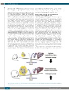
Figure 6. Schematic overview of the proposed mechanism of enhanced survival in the obese mice transplanted with NHD13 bone marrow. MDS: myelodysplastic syndrome; AML: acute myeloid leukemia.
604
haematologica | 2018; 103(4)
