
Page 38 - Haematologica-April 2018
P. 38
F. Pasquier et al. AB
C
DE
F
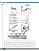
Figure 4. Role of diLeucine motif (diL) loss in the EPOR c.1300dup (p.Gln434Profs*11, EPOR FS) mechanism. (A, B) EPOR internalization was performed with radio- labeled 125I-erythropoietin (EPO). Cells were incubated with 125I-EPO and washed at 4°C to remove unbound ligand. An acidic wash was then performed to separate cell surface–bound from internalized EPO. The radioactivity levels of the (A) supernatant (cell surface–bound EPO) and of (B) the cell pellet (internalized EPO) were determined. Each experiment was performed three times with similar results. (C) To study the potential role of the dileucine motif loss in the EPO hypersensitivity phenotype induced by EPOR FS, Ba/F3 cells were transduced with pMX-HA-huEPOR-IRES-GFP retrovirus to stably express EPOR WT or EPOR STOP receptor with abro- gation of the dileucine motif EPOR WT/diL and EPOR STOP/diL respectively. (D) Proliferation was assessed 48 h after culturing Ba/F3-EPOR cells in the absence or presence of increasing doses of EPO (0.01, 0.02, 0.03, 0.05, 0.1, 0.3, and 1 U/mL) by a WST-1 proliferation assay. Dose-response curves are means expressed in percentages of maximum growth value ± SEM (n = 3 in triplicate). Two-tailed t-test, *P<0.05, **P<0.01, ***P<0.001, ****P<0.0001. (E) Cell-surface expression of the different EPOR was assessed by flow cytometry using PE fluorescence labeling of the extracellular HA-tag. The histogram shows the ratio of mean fluorescence intensity (MFI) of PE-labeled cell-surface EPOR on the respective MFI of GFP. Results are the mean ± SEM of three independent experiments. (F) Effect of EPO con- centration on EPOR signaling. Ba/F3 cells expressing different EPOR constructs were examined by western blotting for the presence and phosphorylation status of various signaling molecules. Cells were serum- and cytokine-starved for 5 h prior to stimulation for 15 min with increasing doses of EPO (0, 0.001, 0.01, 0.1 and 1 U/mL). Expression of β-actin was used as a loading control. One of three independent experiments is presented and fold activation is indicated below.
582
haematologica | 2018; 103(4)
