
Page 24 - Haematologica3
P. 24
J.T. Butler et al.
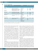
Table 1. Types of extracellular vesicles.
Type of extracellular vesicles
Pseudonym
Exosome
Nanovesicle Nanoparticle
Microvesicle
Microparticle ectosome
Large vesicle
Large oncosome
Apoptotic body
Size (nm)
30-150
50-1000
>1000
>1000
Biogenesis
Early endosomes mature into late endosomes then, through the action of ESCRT, multivesicular bodies are formed containing intraluminal vesicles that fuse with the plasma membrane for release
Direct budding and cleavage of plasma membrane mediated by calcium influx,
and remodeling of the cortical cytoskeleton
Cleavage of large cytoplasmic extensions from cell body
Cytoplasmic fragmentation during programmed cell death
Constituents
Lipid membrane Nucleic acids Proteins Tetraspanins ALIX
TSG101
Lipid membrane
Nucleic acids
Proteins
Tetraspanins SEM
Selection Detection
UC NTA DG Cryo-EM AC TEM
SEC SEM
Reference
[12,22,23, 24, 25, 26]
[12, 27, 28]
[29, 30]
[31, 32]
UC NTA AC Cryo-EM
SEC TEM
Lipid membrane
Nucleic acids
Proteins FACS Organelles
Organized cytoskeleton
Lipid membrane
Nucleic acids FT Proteins FACS Organelles FC Nuclear fragments
Apoptotic markers
CF FM FT FC
CF FM
384
AC: affinity chromatography; CF: centrifugation; Cryo-EM: cryo-electron microscopy; DG: density gradient; ESCRT: endosomal-sorting complex required for transport; FACS: fluo- rescence activated cell sorting; FC: flow cytometry; FM: fluorescence microscopy; FT: filtration; NTA: nanoparticle tracking analysis; SEC: size-exclusion chromatography; SEM: scan- ning electron microscopy; TEM: transmission electron microscopy; UC: ultracentrifugation
mechanisms, upend these homeostatic processes and use EVs to reinforce tumor growth, chemotherapeutic resist- ance, invasion, metastasis and relapse.19-21
EVs can be broadly classified into four subtypes (Table 1) based upon vesicle size and method of cellular release: exosomes (30-150 nm), microvesicles (50-1000 nm), large vesicles (>1000 nm) and apoptotic bodies (>1000 nm).22 It is technically challenging to separate vesicle types, and no standardized method exists to date. Techniques uti- lized for EV purification often rely on size or density.12 However, there is overlap between exosomes and microvesicles in composition and function, and neither size-exclusion chromatography nor ultracentrifugation in density gradients for separation will yield pure popu- lations.22 Moreover, due to overlap between these vesi- cles in size and miRNA carrier function with plasma abundant chylomicrons and very low density lipopro- teins, EV dimension should be considered an arbitrary surrogate metric, and a more biologically informed clas- sification would likely enhance reproducibility in the field, advance their detection and inform treatment strategies.
Exosomes
The biogenesis of exosomes, the smallest type of EV, begins with the inward cleavage of the plasma membrane to form an endosome containing selectively enclosed cytoplasmic components within the lumen. As illustrated in Figure 1C, early endosomes, characterized by the pres- ence of Rab5 protein, undergo maturation into Rab7 con- taining late endosomes which generate multiple intralumi- nal vesicles through the action of tetraspanins and endo- somal sorting complex required for transport (ESCRT)
proteins.25 Together these proteins function to facilitate further inward cleavage and sorting of endosomal con- stituents into discrete intraluminal vesicles. These multi- vesicular bodies, through RAB27- and VPS33b-dependent mechanisms, evade lysosome degradation and fuse with the plasma membrane to release intraluminal vesicles as exosomes.23,24 Through this highly regulated endosomal process of formation, the size of exosomes is relatively constant as compared to the larger types of vesicle. In addition to tetraspanins, proteins ALG-2 interacting-pro- tein X and tumor susceptibility gene 101 (ALIX and TSG101, respectively) are reported to be involved in the endosomal process, and are frequently used as markers for exosomes.12,22 Different cell types can release discrete sub- populations of exosomes, each with different proteomic properties and RNA cargo and divergent membrane pro- tein composition.25,26
Microvesicles
Intermediate-sized EVs are most frequently referred to as microvesicles, ectosomes, or if tumor-derived, oncosomes, which arise via direct budding and cleavage of the plasma. Microvesicles are spherical and span a broad range of sizes, being between 50 nm to 1000 nm in diameter. They are distinguished based on their formation and release, and do not utilize the endosomal/multivesicular body pathway.27 Instead, microvesicles are formed through a process that involves calcium influx and remodeling of the cortical cytoskeleton to release the membrane-enclosed cytosolic cargo.12 Viewed broadly, microvesicles do not appear to be formed in a consistent manner like exosomes. However, when restricted to a specific cell type, microvesicles may form in a uniform manner, as illustrated in one recent study
haematologica | 2018; 103(3)
