
Page 57 - 2020_08-Haematologica-web
P. 57
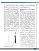
a coagulation inhibitor. Screening for lupus anticocoagu- lant was also negative. Factor VIII coagulant activity (FVIII:C) was 40% (normal range, 50-150%), von Willebrand factor antigen (vWF:Ag) was 18% (normal range. 50-120%), ristocetin co-factor activity (vWF:RCo) was 29% (normal range, 50-150%), and the collagen bind- ing activity (vWF:CB) was 37% (normal range, 50-150%). Following the observation of slightly elevated serum pro- teins (8.8 g/dL; normal range, 6.5-8.0 g/dL), electrophore- sis showed increased concentrations in the beta (β) (2.58 g/dL; normal range, 0.6-0.9 g/dL) and gamma (γ) (2.36; nor- mal range, 0.8-1.4 g/dL) regions, with a double spike at a concentration of 1.67 g/dL (Figure 1). Immunofixation confirmed a double monoclonal component, IgM kappa (k). Immunoglobulin assays showed serum IgG levels of 5.39 g/L (normal range, 7-16 g/L), IgA 0.11 g/L (normal range, 0.7-4 g/L), but very high IgM at 63.7 g/L (normal range, 0.4-2.3 g/L). Bone marrow biopsy detected increased cellularity (90%) that accounted for at least 40% of interstitial cellular aggregates of lymphoid, lymphoplas- macytoid and plasma cells, that at immunohistochemical analysis were positive for CD20, IgM and k light chains but negative for CD5, CD23, D1 cyclin and lambda (λ) light chains. Megakaryocytic and myelo-erythroid lineag- es were represented but depressed. An abdominal ultra- sound showed no hepatosplenomegaly nor lym- phadenopathy. On the basis of these findings a diagnosis of AvWS associated with Waldenstrom macroglobuline- mia was made. The patient underwent a test with desmo- pressin (DDAVP) given subcutaneously at a dose of 0.3 mg/kg in an attempt to increase vWF and FVIII plasma lev- els, but no increase was observed at 1, 2 and 4 hours post injection. Due to the very high serum levels of the IgM monoclonal component, the patient underwent four plas- ma apheretic procedures (each with the removal of 1.5 plasma volume), resulting in a significant reduction in the monoclonal component, and normalization of vWF/FVIII parameters (vWF:Ag 86%, vWF:RCo 77%, vWF:CB 69%, and FVIII:C 84%). The patient was treated with six monthly cycles of bendamustine-rituximab, with a good partial response (IgM 6 g/L, normal hemoglobin and
Figure 1. Example clinical case of acquired von Willebrand syndrome. Patient’s serum protein electrophoresis. The arrows indicate a double spike within the beta (β) and gamma (γ) regions, respectively.
hemostasis test values, complete disappearance of the enlarged lymph nodes). This positive response was main- tained at a 3-year follow up, and a recent bone marrow biopsy showed a normal trilineage hematopoiesis with less than 10% lymphoplasmacytoid cells.
Pathophysiology, clinical presentation, diagnosis
Pathophysiology
In the past, AvWS was considered a very rare hemor- rhagic disease. The more recent discovery of its associa- tion with relatively frequent cardiovascular disorders sug- gests that its prevalence is higher than previously thought.19,20 Unlike acquired hemophilia (another rare acquired bleeding disorder caused by autoantibodies that neutralize FVIII coagulant activity),21-23 the complex patho- physiology of AvWS involves various and different mech- anisms.16-18 Most cases are due to an increased plasma clearance of vWF caused by such mechanisms as antibod- ies, cell adsorption, shear stress or increased proteolysis. At variance with acquired hemophilia, AvWS almost always occurs in association with an underlying disor- der;16-18 besides cardiovascular disorders, those more fre- quently associated are lymphoproliferative disorders [multiple myeloma, chronic lymphocytic leukemia, mon- oclonal gammopathy of undetermined significance (MGUS), Waldenstrom macroglobulinemia], and, less fre- quently, other hematologic malignancies (myeloprolifera- tive neoplasms including essential thrombocythemia, polycythemia vera, primary myelofibrosis, and chronic myeloid leukemia), solid malignancies, and autoimmune disorders (Table 1).16-18,24,25 In a registry of the International Society of Thrombosis and Haemostasis (ISTH) that col- lected data from 211 AvWS cases, lymphoproliferative dis- orders were the most frequent underlying disorder in 48% of cases, whereas myeloproliferative neoplasms and solid tumors accounted for 15% and 5% of cases.16-18 Mohri et al., in a prospective study evaluating 206 patients with a range of hematologic disorders, estimated that AvWS was present in approximately 10% of these patients.25 The dif- ferent underlying disorders lead to AvWS through differ- ent mechanisms. In patients with hypothyroidism, the syndrome is caused by the decreased synthesis of an oth- erwise qualitatively normal vWF, and this can be reversed by l-thyroxine therapy.26,27 In most other cases, the synthe- sis of vWF in megakaryocytes and endothelial cells and its release in circulation are normal, so that AvWS recognizes other mechanistic pathways. In cardiac valvulopathies and left ventricular assist devices, sheering of high-molec- ular-weight (HMW) vWF multimers by mechanical stress or proteolysis induced by ADAMTS 13 (a disintegrin and metalloproteinase with a thrombospondin type 1 motif, member 13) are involved.28-33 In cases associated with plas- ma cell dyscrasias (MGUS and multiple myeloma), as well as in autoimmune diseases such as systemic lupus erythe- matosus, circulating autoantibodies directed against func- tional or non-functional vWF domains have been report- ed.31-33 Antibody binding to vWF leads to the formation of immune complexes that are cleared from the circulation by the reticulo-endothelial system. Antibodies that neu- tralize platelet-related vWF activities (inhibitors) have sel- dom been reported. Finally, a mechanism involving the selective adsorption of HMW multimers on tumor cells leading to their enhanced plasma clearance has been
AvWS focused for hematologists
haematologica | 2020; 105(8)
2033
