
Page 180 - 2020_08-Haematologica-web
P. 180
V.M. Smith et al.
Sensitivity to BH3-mimetics correlated with sequestration of pro-apoptotic BCL-2 proteins
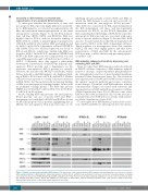
To interrogate whether the interactions of anti- and pro-apoptotic BCL-2 proteins might influence susceptibil- ity to BH3-mimetics, we selected ten representative cell lines and performed immunoprecipitation of the main anti-apoptotic proteins (Figure 3). In the BCL-2-depen- dent cell lines (RIVA, U2932 and OCI-LY1), BIM was highly bound to BCL-2, with no detectable binding of BIM to BCL-XL or MCL-1, despite high protein expression of BCL-XL and MCL-1. In contrast, BIM was highly bound by MCL-1 in the MCL-1-dependent cell lines SUDHL10 and U2946. These two cell lines expressed low levels of BCL-2 and BCL-XL, which may explain why BIM was bound to MCL-1. In the BCL-XL-dependent SUDHL8 and RCK8 cells, BIM expression was comparatively low, and some BIM appeared bound to BCL-XL but not to BCL-2 or MCL-1. Collectively, these data suggest a relationship between the sequestration of BIM by the different anti- apoptotic BCL-2 proteins and a dependency on the respective anti-apoptotic BCL-2 protein for survival. However, the resistant cells OCI-LY3 and Pfeiffer, which did not respond to any BH3-mimetic, also displayed bind- ing of BIM to BCL-2 and/or BCL-XL and MCL-1. Pfeiffer cells have been reported to contain a missense mutation in BIM (S10C), but this mutation did not prevent binding of BIM to its anti-apoptotic binding partners. In line with its published binding profile,28 the BH3-only protein NOXA was exclusively bound by MCL-1 but not by BCL- 2 or BCL-XL in all cell lines.
Besides binding BH3-only proteins, the anti-apoptotic BCL-2 proteins can also sequester BAX and BAK.29 Intriguingly, we found that both BAX and BAK are bound by the anti-apoptotic BCL-2 proteins, highlighting that in DLBCL the anti-apoptotic BCL-2 proteins may act by
inhibiting already partially activated BAX and BAK, in which the BH3-domain is exposed and accessible for interaction with the anti-apoptotic BCL-2 proteins.30 Thus, BAX was sequestered by BCL-2 predominantly in the BCL-2-dependent cell lines, and predominantly sequestered by BCL-XL in the BCL-XL-dependent cell lines, indicating that the binding of BAX by the respective anti-apoptotic BCL-2 protein was associated with sensi- tivity to specific inhibitors (Figure 3). Besides BAX, BAK was also bound by BCL-XL in the BCL-XL-dependent cell lines and by MCL-1 in the MCL-1-dependent cell lines. Taken together, our investigations show that sensitive DLBCL cell lines were highly primed and that direct sequestration of BAX and BAK by the anti-apoptotic BCL-2 proteins could be the last step preventing apopto- sis in these cells.
BH3-mimetics induced cell death by displacing and activating BAX and BAK
Next, we asked how BH3-mimetics induced cell death in DLBCL cell lines. Exposure to BH3-mimetics induced caspase-3 cleavage, caspase-dependent phosphatidylser- ine externalization and loss of mitochondrial membrane potential (Online Supplementary Figure S4). The activation and oligomerization of BAX and/or BAK are key events in the intrinsic apoptotic pathway and require conforma- tional changes. Treatment with BH3-mimetics induced conformational changes associated with activation and oligomerization of BAX and BAK in all sensitive cell lines (Online Supplementary Figure S5A-C). Of note, some active BAK was detectable in untreated cells, but the amount of constitutively active BAK did not correlate with sensitiv- ity (Online Supplementary Figure S5D).
To investigate how BH3-mimetics induced the activa- tion of BAX and BAK we interrogated how the interac-
Figure 3. Priming correlates with sensitivity to BH3-mimetics. The interaction of anti- and pro-apoptotic BCL-2 proteins was investigated in a selection of ten cell lines with varying sensitivities to BH3-mimetics. Immunoprecipitation of BCL-2, BCL-XL and MCL-1 was performed in untreated cell lysates followed by analysis of binding of pro-apoptotic BCL-2 proteins (BIM, NOXA, BAX and BAK) using Western blotting. Protein G beads without primary antibody were used to control for unspecific bind- ing. Staining with BCL-2, BCL-XL, MCL-1 and GAPDH was performed to demonstrate efficient immunoprecipitation and equal protein loading, respectively. Representative western blots of two independent experiments are shown.
2156
haematologica | 2020; 105(8)
