
Page 176 - 2020_08-Haematologica-web
P. 176
V.M. Smith et al.
Results
BCL-2, BCL-XL and MCL-1 are important therapeutic targets in diffuse large B-cell lymphoma
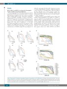
To investigate the roles of the main anti-apoptotic BCL- 2 proteins in DLBCL, we assessed the effects of selective BH3-mimetics in DLBCL cells. We focused on commer- cially available inhibitors that target BCL-2 (ABT-199), BCL-XL (A1331852, A1155463) or MCL-1 (A1210477,21 S63845). Primary cells isolated from patients’ tissues were exposed to different concentrations of BH3-mimetics before analysis of cell viability using a CellTiterGlo Assay (Figure 1A). The direct comparison of ABT-199, A1331852 and S63845 revealed that the response to BH3- mimetics was highly heterogeneous, with three of seven samples (#1, #2, and #3) responding to low nanomolar concentrations of S63845, sample #4 responding best to
ABT-199, and samples #5, #6 and #7 being more resistant to all three BH3-mimetics. Notably, sample #3 displayed a better response to A1331852 than to ABT-199, indicat- ing that although none of these primary samples dis- played the highest sensitivity to A1331852, all three main anti-apoptotic BCL-2 proteins may be relevant therapeu- tic targets in DLBCL.
As primary patient-derived DLBCL cells are limited and freshly isolated malignant B cells rapidly lose viability ex vivo, we continued our investigations in a panel of 18 DLBCL cell lines comprising the main subtypes of DLBCL defined by gene expression profiling.22 namely activated B-cell, germinal center and primary mediastinal B-cell lymphoma-like cells (Table 1). In addition, based on their mutation/translocation signature derived from public databases, we characterized the cell lines according to their genetic drivers into MCD (MYD88 and CD79b
AB
C
D
Figure 1. Diffuse large B-cell lymphoma cells display a heterogeneous sensitivity to selective BH3-mimetics. (A) Primary cells isolated from patients’ tissues were incubated with different concentrations of ABT-199, A1331852 or S63845 for 24 h before analysis of cell viability using CellTiterGlo. Experiments were performed in triplicate and data shown are the mean and standard deviation (SD) for each individual sample (n=7). (B-D) Diffuse large B-cell lymphoma cell lines were exposed to different concentrations of ABT-199 (B), A1331852 (C) or S63845 (D) before analysis of cell viability using CellTiterGlo at 72 h. Data shown are the mean and SD (n=4-6). Half maximal effective concentration (EC50) values, as displayed in Table 1, are indicated for highly sensitive cell lines.
2152
haematologica | 2020; 105(8)
