
Page 113 - 2020_08-Haematologica-web
P. 113
PDGFRb regulation in murine myelofibrosis
marked spindle-shaped stromal cells in early and overt fibrotic bone marrow of Gata-1low mice. Staining of the ligand PDGF-A showed expression in a wide variety of different hematopoietic cells, whereas PDGF-B mainly derived from megakaryocytes, suggesting a paracrine effect of PDGF-B on PDGFRb in bone marrow fibrosis.
Whereas megakaryocyte dysplasia and proliferation is a defining feature of PMF, fibroblast proliferation leading to a progressive fibrosis is the key pathological aspect of PMF. Given the distinct expression of PDGFRb in stromal cells of early fibrotic and fibrotic bone marrow, we fur- ther concentrated on the dynamics of PDGFRb and its lig- and PDGF-B.
Ligand-activation and regulation of PDGFRb during the development of myelofibrosis
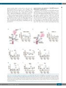
Figure 5. Interaction analysis of platelet-derived growth factor receptor b (PDGFRβ) with PDGF-B and PDGFRb tyrosine phosphorylation in the bone marrow of Gata-1low mice at 5 months (5 M), 10 months (10 M) and 15 months (15 M) of age. (A) Illustration of a proximity ligation assay (PLA) for the analysis of PDGFRb– PDGF-B interaction. (B) Quantitative analysis of PDGFRb–PDGF-B interaction by PLA in the bone marrow of Gata-1low mice and age-matched wild-type (WT) controls. (C) Illustration of a PLA for the analysis of PDGFRb tyrosine phosphorylation. (D) Quantitative analysis of PDGFRb tyrosine phosphorylation by PLA in the bone marrow of Gata-1low mice and age-matched WT controls, n=6 mice per group, 2670-9001 nucleated cells per mouse were analyzed for the presence of rolling circle products (RCP). (E) qPCR analyses of Ptpn1, (F) Ptpn2, (G) Ptpn6, (H) Ptpn11, (I) Ptpn12 and (J) Ptprj, n=7 mice per group, *P≤0.05, **P≤0.01, ***P≤0.001 versus the con- trol group by Student t-test.
The increased protein expression of PDGFRb and its lig- and PDGF-B in overt myelofibrosis prompted us to inves- tigate the interaction of both signaling components in situ. To analyze PDGFRb–PDGF-B binding, we performed a PLA using combined primary antibodies detecting PDGFRb and PDGF-B (Figure 5A). The assay allows for the detection and quantification of signals and showed an increased interaction of receptor and ligand in overt fibrot- ic bone marrow of 15-month-old Gata-1low mice (Figure 5B, original PLA images are shown in Online Supplementary Figures S5). This is in accordance with enhanced protein
ABCD
EFG
HIJ
haematologica | 2020; 105(8)
2089
