
Page 111 - 2020_08-Haematologica-web
P. 111
PDGFRb regulation in murine myelofibrosis
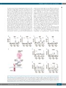
revealed a major increase in ligand gene expression at the early fibrotic stage (Figure 3C and D). We again observed a decrease in Pdgfa gene expression in pre-fibrotic bone marrow, whereas Pdgfa gene expression was increased in early and overt fibrotic bone marrow. Pdgfb expression was significantly up-regulated only in early fibrotic bone marrow and remained at nearly baseline level in pre- fibrotic and overt fibrotic bone marrow of Gata-1low mice.
To validate the gene expression data, we further ana- lyzed protein expression using an in situ proximity liga- tion assay (PLA) in a single recognition approach (Figure 3E). By this method, two oligonucleotide-coupled sec- ondary antibodies (PLA probes) detect a single primary antibody. Through ligation, oligonucleotides are joined to a circle when in close proximity and serve as template for polymerization. A polymerase replicates the DNA circles and a concatemeric product is generated. Fluorescently- labeled nucleotides enable the detection of a rolling circle product (RCP), which can be visualized and quantified as a distinct fluorescent dot.17,18 Corresponding negative con- trols, positive controls, and results from the quantitative
analyses by proximity ligation assay (PLA) in comparison to quantified protein expression data acquired from mul- tiplex staining are shown in Online Supplementary Figures S1-S3. Although we observed a heterogenic protein expression in Gata-1low mice, there was a steady increase in PDGF receptor protein expression during the develop- ment of myelofibrosis (Figure 3F and G; original PLA images are shown in Online Supplementary Figures S4). When analyzing PDGF-A and PDGF-B protein expression by single recognition PLA, we again observed high het- erogeneity among age-matched Gata-1low mice. We did not detect a significant increase in PDGF-A expression, while PDGF-B protein expression was significantly increased in overt fibrotic bone marrow of 15-month-old Gata-1low mice (Figure 3H and I, original PLA images are shown in Online Supplementary Figures S4).
To visualize the cell type-specific expression of the PDGF signaling components, we performed multiplexed staining of PDGF signaling components in the bone mar- row (Figure 4). We observed PDGFRa expression pre- dominantly in megakaryocytes, whereas PDGFRb
ABCD
EFG
HI
Figure 3. Expression of platelet-derived growth factors (PDGF) and their receptors in the bone marrow of Gata-1low mice at 5 months (5 M), 10 months (10 M) and 15 months (15 M) of age. (A) Quantitative polymerase chain reaction (qPCR) analyses of Pdgfra, (B) Pdgfrb, (C) Pdgfa, and (D) Pdgfb in the bone marrow of Gata-1low mice and age-matched wild-type (WT) controls. n=7 mice per group. (E) Illustration of a single recognition proximity ligation assay (PLA) as a sensitive means to quantify protein expression in situ. (F) Quantitative analyses of PDGFRa, (G) PDGFRb, (H) PDGF-A, and (I) PDGF-B protein expression by single recognition PLA in the bone marrow of Gata-1low mice and age-matched WT controls. n=6 mice per group. 3170-7935 nucleated cells per mouse were analyzed for the presence of rolling circle products (RCP). *P≤0.05, **P≤0.01, ***P≤0.001 versus the control group by Student t-test.
haematologica | 2020; 105(8)
2087
