
Page 112 - 2020_08-Haematologica-web
P. 112
F. Kramer et al.
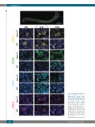
Figure 4. Multiplex staining of platelet-derived growth factors (PDGF) and their receptors in the bone marrow of Gata-1low mice at 5 months (5 M), 10 months (10 M) and 15 months (15 M) of age. Representative images showing femoral bone marrow of Gata-1low mice and wild-type (WT) control mice stained for PDGF receptor a (PDGFRa, yellow), PDGF receptor b (PDGFRb, green), PDGF-A (cyan) and PDGF-B (magenta). 10 images at 40x magnification were acquired within the femoral bone marrow for each group. Nuclei were counterstained with DAPI (blue), scale bars in lower panels=20 mm.
2088
haematologica | 2020; 105(8)
