
Page 98 - 2020_07-Haematologica-web
P. 98
C. Casu et al.
AB
CD
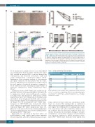
Figure 4. The iron-restrictive effect of minihepcidin improved ineffective erythropoiesis in Hbbth1/th2-BMC mice. Minihepcidin improved (A) red blood cell (RBC) morphology and (B) RBC lifespan [statistics are determined comparing animals treated with vehicle (V) vs. high-dose minihepcidin (MH_H)]. (C) Minihepcidin also improved erythropoiesis in the bone marrow (BM) and spleen of Hbbth1/th2-BMC mice. (D, E) Flow activated cell sort- ing analysis of the percentage of BM (D) and splenic (E) erythroid cells using CD44+ and Ter119+ cells (n=4-5 animals per group). Results are presented as means ± standard deviation: ****P≤0.001, **P≤0.01, *P≤0.05.
blood transfusion in animals treated or not with MH_H (see experimental design in Online Supplementary Figure S4B) resulted in increased RBC count and hemoglobin concentration, and decreased reticulocyte count and serum erythropoietin concentration (Figure 6A-D). Furthermore, flow cytometric analysis of bone marrow and splenic erythroid cells demonstrated that the combi- nation of MH_H and blood transfusion further reduced the total number of erythroid progenitors compared to blood transfusion alone, indicating an improvement of ineffective erythropoiesis (Online Supplementary Figure S6A-C).
Transfusion alone in Hbbth1/th2BMC mice resulted in sig- nificantly increased serum hepcidin (Figure 7A), likely due to suppression of both serum erythropoietin concentra- tion (Figure 6D) and endogenous erythropoiesis (Online Supplementary Figure S6). Administration of MH_H (with and without blood transfusion) had little effect on trans- ferrin saturation (Figure 7B), but improved serum iron lev- els (Figure 7C) in non-transfused Hbbth1/th2BMC mice. Compared to Hbbth1/th2BMC mice treated with vehicle alone, transfusion significantly decreased liver iron con- centration (Figure 7D, Online Supplementary Figure S7), likely due to the increased levels in serum hepcidin (Figure 7A), but no further decrease was observed in MH_H-treat- ed transfused Hbbth1/th2BMC mice.
Appreciable iron deposition in the heart makes our model helpful to study a pathological feature extremely relevant in patients affected by thalassemia major. In par-
Table 1. Serum erythroferrone measurements. Erythroferrone ng/mL
WT Hbbth1/th2-V
Hbbth1/th2_MH_H****
1.0 9.6 4.3
LLOQ 17.1 8.5
LLOQ 13.0 7.0
2.2 14.3 6.8
LLOQ 15.2 7.7
LLOQ 9.6 4.3
2.4 17.1 8.5
Serum erythroferrone levels in wildtype (WT) and thalassemic animals, treated with vehicle (V) or a high dose of minihepcidin (MH_H). A statistically significant differ- encewasobservedcomparingHbbth1/th2 BMCvehicle-treatedvs.Hbbth1/th2 BMCMH_H- treated animals (****P≤0.001).The lower limit of quantification (LLOQ) in wildtype animals was 0.25 ng/mL.
ticular, when we looked at the iron concentration in the heart, we observed that minihepcidins in combination with a transfusion regimen significantly reduced iron con- tent (Figure 7E). Furthermore, as minihepicidins enable iron sequestration and reduce ineffective erythropoiesis, we postulate that the decreased erythroid mass also reduces the amount of iron utilized, leading to a relative normalization of transferrin saturation and parenchymal iron deposition. Furthermore, MH_H treatment in trans- fused Hbbth1/th2BMC mice decreased total spleen iron
1840
haematologica | 2020; 105(7)
