
Page 96 - 2020_07-Haematologica-web
P. 96
C. Casu et al.
[high dose (MH_H)] 2 months after transplantation. The experimental design is shown in Online Supplementary Figure 4A, B. The duration of the treatment was selected based on the findings of our previous pharmacokinetic studies.28 Compared to controls (V- vehicle), administra- tion of minihepcidins improved hematologic parameters in a dose-dependent manner. Using the lowest dose, we observed a trend of improved parameters, with the improvement reaching statistical significance with the highest dose. RBC count and hemoglobin concentration were statistically significantly improved in animals treat- ed with the high dose (Figure 3A, B). Similarly, reticulo- cyte count and splenomegaly decreased more in MH_H- treated Hbbth1/th2BMC mice (Figure 3C, D). We then focused only on the highest dose. Minihepcidin adminis- tration also decreased hemichrome formation (Figure 3E) and reactive oxygen species production (Figure 3F). Accordingly, RBC morphology (Figure 4A) and lifespan (Figure 4B) improved in MH_H-treated mice, relative to vehicle-treated Hbbth1/th2BMC mice. Flow cytometric analysis of bone marrow and spleen samples (Figure 4C) demonstrated improved ineffective erythropoiesis in minihepcidin-treated Hbbth1/th2BMC mice as the percent- age (Figure 4D, E) of erythroid progenitor cells decreased compared to that of mature RBC.
Administration of minihepcidins ameliorated iron overload in untransfused Hbbth1/th2BMC mice
As erythropoiesis improved in Hbbth1/th2BMC MH_H- treated mice, we investigated whether minihepcidins had a beneficial effect on endogenous hepcidin synthesis and iron metabolism. Hbbth1/th2BMC mice treated with vehicle demonstrated a significant increase in serum erythrofer- rone levels compared to WT animals, but a reduction in these values when treated with MH_H (Table 1, Figure 5A). Endogenous serum hepcidin concentrations were dif- ferent between untreated and treated animals (Figure 5B), but no significant differences were observed in transferrin saturation levels (Figure 5C). However, serum iron levels decreased significantly in MH_H-treated Hbbth1/th2BMC mice (Figure 5D). Moreover, Hbbth1/th2BMC MH_H-treated mice showed significant reductions of iron by ~33% and ~77% in the liver and spleen, respectively, but not in the kidney (tissue iron content in the kidney not shown). (Figure 5E, F and Online Supplementary Figure S5).
Minihepcidin treatment ameliorated ineffective erythropoiesis, reversed splenomegaly, and reduced serum iron and heart iron concentration in transfused Hbbth1/th2BMC mice
Compared to Hbbth1/th2BMC mice treated with vehicle,
ABC
DE
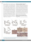
Figure 2. Complete blood count analysis of animals affected by β-thalassemia intermedia or major. (A) Red blood cell (RBC) number, (B) hemoglobin (Hb) levels, (C) reticulocytes (RETIC) count and (D) spleen weight. Bars represent standard deviation. ****P≤0.001. (E) RBC morphology (shown by Giemsa staining of peripheral blood smears) of wildtype (WT), β-thalassemia intermedia and Hbbth1/th2-BMC mice.
1838
haematologica | 2020; 105(7)
