
Page 94 - 2020_07-Haematologica-web
P. 94
C. Casu et al.
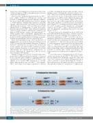
of 3.7 Kb containing the β-major gene and 2 Kb of the 5’ flanking region, including the promoter (Figure 1A).34 Hbbth2/+ mice were created by inserting a neomycin-resis- tant cassette into exon 2 of the β-major gene such that heterozygotes are mildly anemic while homozygotes die perinatally due to severe anemia (Figure 1B).35 Hbbth3/+ mice have one copy of the normal β-globin cluster and an allele with a deletion of both the β-major and β-minor genes (Figure 1C), resulting in moderate anemia that is not severe enough to require transfusion, a phenotype similar to that of Hbbth1/th1 mice.36,37 Homozygous Hbbth3/th3 mice die perinatally, preventing their use as an adult model of TDT.36
We previously used a transplant model in which fetal liver cells from E13.5-15.5 day Hbbth3/th3 embryos are trans- planted into irradiated wildtype (WT) syngeneic mice.36-38 Successful engraftment of Hbbth3/th3 fetal liver cells led to ineffective erythropoiesis and severe anemia resulting in death 3 months after transplantation if the animals were not transfused.16,38,39 This and other models were utilized to study dysregulated iron metabolism in β-thalassemia major.16,35-39 However, Hbbth3/th3 mice are characterized by such low hemoglobin and RBC production that they make testing drugs, such as minihepcidins that have the potential to modify RBC quality and lifespan and improve ineffective erythropoiesis, complex if not impos- sible.
To assess the efficacy of minihepcidins in TDT, we gen- erated a new mouse model (Hbbth1/th2) that closely resem- bles the human TDT phenotype (Figure 1D). Our aim was to use combinations of already existing mutations in order to generate a model intermediate in severity to those already in use, in which some RBC are produced although their synthesis is insufficient to support long-
ABC
logical way of excreting the iron recycled from these cells, continuous infusion of RBC is the primary reason for iron overload in TDT patients.18,21,22
Mouse models of β-thalassemia intermedia (e.g. Hbbth3/+ mice) exhibit ineffective erythropoiesis, anemia and reduced or inappropriately normal hepcidin synthesis, but do not require RBC transfusion for survival, similarly to NTDT patients. Minihepcidins function as hepcidin agonists, target ferroportin, and reduce iron absorption and transferrin saturation.23,24 We and others showed that administration of minihepcidins or agents that induce hepcidin expression in Hbbth3/+ mice decreased transferrin saturation, heme synthesis, hemichrome formation, and improved RBC lifespan, anemia, and splenomegaly.17,25-29 Taken together, these experiments demonstrated the potential benefits of minihepcidins in NTDT. However, it is unclear whether minihepcidins would improve anemia, transfusion requirements, and iron overload in TDT.
Based on the pathophysiology of TDT and the effect of minihepcidins on iron metabolism and erythropoiesis in NTDT, we speculate that minihepcidins may: (i) improve ineffective erythropoiesis; (ii) increase RBC lifespan and reverse anemia; (iii) decrease RBC transfusion require- ments (decrease frequency of transfusion); (iv) reverse splenomegaly and extramedullary erythropoiesis; (v) decrease indications for splenectomy; and (vi) reverse iron overload in TDT patients.
Multiple existing mouse models of β-thalassemia inter- media harbor different mutations leading to decreased mouse β-globin genes synthesis, triggering ineffective erythropoiesis and anemia (Figure 1A-C). However, some animals do not require RBC transfusion for survival, while others produce very few RBC.30-33 For example, Hbbth1/th1 mice carry a homozygous spontaneous deletion
D
Figure 1. Genetic makeup of established mouse models of β-thalassemia intermedia and a new model of ß-thalassemia major. (A-C) Mouse models of β-tha- lassemia Intermedia: (A) Hbbth1/th1, (B) Hbbth2/+, (C) Hbbth3/+ and (D) a new mouse model of β-thalassemia major: Hbbth1/th2. The mouse β-globin locus is represented in the 5’ to 3’ orientation; for simplicity, only the β-globin genes are indicated; not in scale. LCR: β-globin locus control region; βma: β-globin major gene; βmi: β-globin minor gene; N: neomycin gene. Dotted lines represent DNA deletions.
1836
haematologica | 2020; 105(7)
