
Page 40 - 2020_07-Haematologica-web
P. 40
P.A. Egan et al.
to the BM plasma cells. Overall, the ability to carry out iFISH or molecular testing was compromized in most instances by inadequate sample and/or myeloma cell numbers.
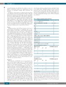
A summary of presentation, treatment and survival data from all papers reviewed is presented in Table 2. Although limited by variations in both the approach and incomplete data in the original manuscripts, this analysis confirms the bias towards a lower M:F ratio, and more frequent λ light chain restriction than in ND-MM without CNS involve- ment. Furthermore, CNS relapse 26 months following MM diagnosis is in keeping with the duration generally quoted. Because of incomplete data, definitive treatment analysis preceding and following CNS-MM relapse could not be ascertained. However, within these limitations, summary treatment data are annotated in Table 2.
Cause
radiotherapy, intrathecal chemotherapy, and IMiD.15 Majd et al. studied nine CNS-MM patients treated between 2008-2013 and observed that the three longest survivors received stem cell transplant after CNS involvement was detected.25 Interestingly, none of these nine patients was receiving maintenance therapy before CNS involvement was detected.25
Table 2. Analysis of data from studies referenced.
Parameter
% detected at MM diagnosis
Months from MM diagnosis to CNS-MM % male
Age
%IgG
Mean of all studies (range)
16
26 (0 - 216) 57
57
38
26
Multiple myeloma with CNS involvement develops via
hematogenous dissemination of malignant cells or con-
tiguous spread of the tumor, often associated with PCL
and cranial plasmacytoma, respectively.1,15 Although it has
been suggested that invasion of the CNS is enabled by
treatment of MM with immunomodulatory drugs (IMiD),
with a report of an MM patient receiving lenalidomide
prior to CNS-MM progression,17 this is not robust evi-
dence. Data for EMD in general suggest that escape from
the BM is enabled by mutations to tumor suppressor
genes such as TP53, oncogenes such as RAS, and altered
expression of adhesion molecules, as outlined above.18-21
These genetic changes may enable proliferation independ-
ent of stimuli provided by the BM environment.
Furthermore, recent studies do not support a causal link
between modern MM treatment and subsequent EMD
which may rather be a consequence of longer survival of
patients treated with novel agents.2,21-23 Additionally, recent
increases in EMD prevalence have been seen at MM diag-
nosis as well as post treatment, and therefore may be due
to improved detection.2 In another study, the only risk fac-
tor for an extramedullary relapse following autologous
stem cell transplant (SCT) was EMD at MM diagnosis.5
Further weak evidence for a causal relationship between
loss of neural cell adhesion molecule (NCAM) (CD56) and
CNS-MM, which has a role in cell-cell adhesion, is pre-
sented in our own data (Table 1). None of the above
%IgA
%IgD 4 % biclonal 5 % light chain only 21 % lambda 50
iFISH on CSF (compared BM at MM Dx)
13q loss 33% (38%)
17p loss 14% (9%)
1q gain 10% (17%)
t(4;14) 14% (9%)
t(11;14)14% (5%)
Courses of MM treatment before CNS-MM OS from CNS-MM diagnosis (Months) MM treatment
2.2
4.5
CNS-MM median OS
√
CNS-MM treatment
The majority of CNS-MM cases are in patients who
have received MM therapy prior to CNS involvement
(Table 2) and whose survival is generally short and may
depend on subsequent treatment.6,8,15,24 In a recent retro- √√ spective study of 172 CNS-MM patients, Jurczyszyn et al.
√
SCT
√ √
10.9 3.0 √ 4.0 1.6
CNS-MM median OS
IMiD PI SCT XRT
√√ 2.6 √ √ 6.0 √√√ 3.5
Prognosis
IMiD PI
XRT
√ 4.7
found the median overall survival (OS) from the onset of CNS involvement to be seven months; multivariate analy- sis revealed that receiving MM therapy before CNS involvement, and having >1 cytogenetic marker of poor prognosis, were risk factors that reduced median OS from 25 months to 5.5 months when either was present, and to two months with both present.8 Jurczyszyn et al. also showed a median OS of 12 months in patients who received systemic therapy following CNS-MM diagnosis.8 Similarly, Chen et al. analyzed records for 37 patients treated between 1999-2010 and found a group of nine longer survivors with a median OS of 17.1 months from CNS-MM diagnosis, who were typically treated with
√
None of the above
√ 7.3 5.8 √ 2.0 √ 6.0 √ 9.0 1.0
√√ √
5.1
Summary, where data are available. Means and medians were weighted according to study size and used to calculate an overall mean. MM: multiple myeloma; CNS-MM: multiple myeloma with central nervous system (CNS) involvement; OS: overall sur- vival. Cerebrospinal fluid (CSF) interphase fluorescence in situ hybridization (iFISH) data from 21 cases (3 studies) compared to that from 64 cases (12 studies) at diagno- sis (Dx) of multiple myeloma (MM Dx).Treatment data obtained from 123 cases of CNS-MM. Prior to CNS-MM diagnosis, 36% of patients received one or more stem cell transplants (SCT); 27% were treated with one or more immunomodulatory drugs (IMiD); 24% received a proteasome inhibitor (PI); and 9% received radiotherapy (XRT). BM: bone marrow.
1782
haematologica | 2020; 105(7)
