
Page 42 - 2020_07-Haematologica-web
P. 42
P.A. Egan et al.
less frequently, seizures, vomiting, cranial nerve palsy, lethargy, fever, convulsion, vertigo, hearing loss and incon- tinence.1,8 When such symptoms are seen in MM patients, the ensuing investigations employ imaging, cytological and/or cytometric techniques. The suggested approach to diagnosis of CNS-MM is shown in Figure 1.
Cytological techniques can detect atypical plasma cells and flow cytometry can detect monoclonal CD38/CD138 expressing cells in CSF in approximately 90% of CNS- MM cases, thus confirming the disease.8,41 CSF cytology and flow cytometry are both particularly useful since the former can employ immunocytochemistry to identify unknown tumors,42 and the latter can be used to distin- guish the clonal plasma cells found in MM from polyclon- al plasma cells present in CSF in other conditions.43 Furthermore, the presence of a paraprotein, including clonal free light chains (FLC), in CSF obtained from a clean lumbar puncture, can be diagnostic. Minute or unde- tectable concentrations of paraprotein in the parallel analysis of serum is strong evidence that monoclonal immunoprotein detected in CSF originates from plasma cells in the CNS rather than BM.
In the study of 172 CNS-MM patients by Jurczyszyn et al., magnetic resonance imaging (MRI) of the brain and/or spine showed evidence of CNS involvement in 93% of cases, while computed tomography (CT) scans showed evidence in 81%.8 In the patients who underwent imag- ing, leptomeningeal involvement was found in over half, intracranial mass in approximately half, and both in
approximately 20%.8 Fluorescence in situ hybridization can reveal EMD and is therefore potentially useful for detection of CNS-MM.44,45 Diagnosis of CNS-MM is con- firmed using imaging and by detection of monoclonal immunoprotein and/or clonal plasma cells in CSF (Figure 2), with the last of these especially useful for lep- tomeningeal involvement.25,35 Imaging techniques are effective in most cases, although studies estimate a 10% false negative rate.8 Detection of plasma cells in CSF pro- vides strong evidence of CNS-MM, although these can be absent when infiltration of parenchymal CNS has occurred.8,46
Treatment of multiple myeloma with CNS involvement: current approaches and future directions
The optimal approach to treatment of CNS-MM is not currently known. The relatively small numbers of patients presenting with this complication means that there is no high quality, prospective clinical trial data to inform an evidence-based approach to therapy. The current approach mirrors those treatment modalities used in lym- phoproliferative disease infiltrating the CNS, namely, sys- temic therapy, intrathecal (IT) therapy, and CNS irradia- tion, often in combination.
Systemic therapy
Drug therapies successfully employed in MM might be ineffective in CNS-MM due to: tumor resistance after pre- vious therapy,8 because they require interaction with the
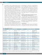
Table 3. Studies considered in this review.
Reference
Nieuwenhuizen L and Biesma DH. 20081
Varga G et al. 20186 Jurczyszyn A et al. 20168 Paludo J et al. 20169
Study dates CNS-MM
1968-2007 109*
2007-2017 13
1995-2014 172
1998-2014 29
Topic of study
Literature review – diagnosis and treatment
Imaging, CSF analysis,
treatment, survival
Multicenter study of pathology, imaging and survival
Plasma cell detection in CSF
CNS-MM and novel agents
Risk markers including
cytogenetic
CSF protein, intrathecal therapy Diagnosis and treatment Treatment and survival
CNS-MM concurrent with PML Cytogenetics
Cytogenetics
Characterization
Intracranial EMD
and novel therapies Brazilian center Large-scale MM study
Flow cytometry
Reference
Fassas AB et al. 200232
Chang H et al. 200533
Liu XJ et al. 201534
Marini A et al. 201435
Lopes AC et al. 201736 Kaplan JG et al. 199040
Mendez CE et al. 201046 Fukunaga H et al. 201744 Bommer M et al. 201841
Ren H et al. 201742
Riley JM et al. 201158 Katodritou E et al. 201552 Vicari P et al. 200351 Mussetti A et al. 201354
Badros A et al. 201755
Kauffmann G et al. 201759
Marron TU et al. 201582
Study dates CNS-MM
1990-2002 18*
2005 8
2015 1
2014 1
2017 1
1990 63
2010 1 2017 1 2017 16
2017 2
2011 1 2000-2013 31 2003* 54
2009-2013 1
2008-2016 2
2017 1
2011-2013 9
Topic of study
Features associated with CNS-MM including cytogenetic
CSF plasma cell, CD56
Case description
Flow cytometry for rapid
diagnosis, CD56
CD56+ CNS infiltration Presentation and cytology
Case study with dural involvement FDG-PET
Cytology, flow cytometry and iFISH for diagnosis
CSF cytology for diagnosis Radiotherapy
Treatment with novel agents Thalidomide
Pomalidomide
Marizomib
Proton therapy
FLC measurement in CSF
Gangatharan SA et al. 201210 2001-2010 7
Fassas AB et al. 200411 Lee D et al. 201312
Abdallah AO et al. 201413 Chen CI et al. 201315
Ruiz-Heredia Y et al. 201817 Chang H et al. 200418 Chang WJ et al. 201424 Majd N et al. 201625 Gozzetti A et al. 201226
Dias A et al. 201827
Kyle RA et al. 200328
Marchesi F et al. 201629
1990-2004 25**
2000-2011 17 1996-2012 35 1999-2010 37
2018 1 2000-2003 9 2006-2010 8 1998-2012 9 2000-2010 0
2008-2016 3 1985-1998 0***
2016 4
*Nieuwenhuizen et al. (2008)1 included 18 cases from Fassas et al. (2002)32 and 54 cases from Vicari et al. (2003).51 **Fassas et al. (2004) 11 includes 18 cases from Fassas et al. (2002).32 ***Multiple myeloma cases only. CNS: central nervous system; MM: multiple myeloma; CNS-MM: multiple myeloma with CNS involvement; CSF: cerebrospinal fluid; PML: progressive multifocal leukoen- cephalopathy; FDG-PET: fluorodeoxyglucose positron-emission tomography; FLC: serum free light chain; iFISH: interphase fluorescence in situ hybridization.
1784
haematologica | 2020; 105(7)
