
Page 39 - 2020_07-Haematologica-web
P. 39
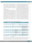
Table 1. Regional hematologic malignancy diagnostic service data (SO’C, 2019, unpublished data).
CNS-MM
diagnosis.7-10 Infiltration of the CNS or meninges is rarer in myeloma than in most other hematologic malignancies, affecting well under 1% of patients, and carries a very poor prognosis with reported median overall survival (OS) of seven months or less following its diagnosis.8-13 However, intracerebral plasmacytomas that develop from osseous lesions of the cranium can be treated successfully with radiation, unlike the more serious myelomatous meningitis.14
Incidence and prevalence
The reported median age of onset of CNS-MM is often younger (50-60-year old age group) than the usual median age of approximately 70 years for MM diagnosis, with up to 20-25% of cases discovered at the initial myeloma diag- nosis.8,15 However, age at presentation varies between stud- ies, including that of our own data (Table 1), suggesting CNS-MM may be underdiagnosed in older patients. CNS- MM can arise at any stage of MM, and although previous studies suggest a bias towards later stage disease,1 a recent large-scale retrospective study did not find an association with MM clinical stage.8 The improved OS of MM patients is expected to lead to an increased incidence of EMD and CNS-MM, possibly due to the extra time avail- able for mutations in residual, drug-resistant tumor cells following treatments, that alter expression of adhesion molecules, oncogenes and tumor suppressor genes.14 Furthermore, there may also be an increase in the time
from MM diagnosis to CNS involvement due to the effec- tiveness of high-dose chemotherapy and treatment using novel agents.10 Indeed, patients have often had several lines of treatment by the time CNS-MM is diagnosed.8,16
In our own experience in a regional hematologic malig- nancy diagnosis center (HMDS, Leeds, UK) over a 15-year period (December 2003-March 2019), ten cases (6 female, 4 male) of CNS-MM were identified (SO’C, 2019, unpub- lished data). Two of these were at MM presentation, whilst the remainder occurred 6-108 months following MM diag- nosis (Table 1). The incidence was well under 1% overall (5,238 cases of MM were investigated at HMDS during this period). A higher incidence of female (F) to male (M), and lambda (λ)-restricted to kappa (k)-restricted, patients, to that found in newly diagnosed MM (ND-MM), was noted. Although absence of CD56 expression was more frequent (4 out of 10 cases) than seen in ND-MM, and one case showed rearranged immunoglobulin heavy chain (IGH), and one loss of 1p with gain of 1q, none of these parameters, including immunophenotypic or acquired cytogenetic aberrations, was seen in adequate numbers to be suggestive of significant association with CNS involve- ment. Furthermore, bone marrow (BM) interphase fluo- rescence in situ hybridization (iFISH) was not available in the earlier cases, so association of CNS-MM with cytoge- netic aberrations predisposing to its development cannot be reported due to small sample size. In all cases, the immunophenotype of the CNS plasma cells was identical
Case n.
1
2 3
4
5
6
7
8
9
Gender
F
F M
F
M
F
M
F
M
Age at CNS-MM
76
89 71
90
55
77
76
57
70
MM to CNS- CNS CD56 MM (months) status
20 CD56+/-
BM FISH
*
* *
*
*
Deletion TP53, monosomy 13, IGH-MAF translocation
Insufficient sample
CNS FISH
No
No
IGH rearranged
No
No
No
No
No
No
1q21.3 gain, 1p32.3 loss
Additional comments
Insufficient CSF sample for full FISH panel
Patient presented with CSN disease (limb weakness and cranial nerve palsy). BM aspirate not received
Patient presented with CNS disease (cranial nerve palsy), BM requested after CSF sample report
Bone plasmacytoma, myeloma diagnosed on BM; plasma cell leukemia 2 months
prior to CNS disease
Multiple plasmacytomas, myeloma
diagnosed on BM
Concurrent plasma cell leukemia and CNS disease
Identical iFISH cytogenetic abnormalities as at presentation despite 108-month separation
n/a
18 15
0
0
6
41
28
28
CD56- CD56-
CD56-
CD56++
CD56+/-
CD56++
CD56+
CD56-
CD56+
4/10 CD56-
2/10 CD56wk
IGH-FGFR3 translocation, 1q21 gain, 13q loss
Hyperdiploid (Chr 5, 9, 15)
1q21.3 gain, 1p32.3 loss
10 F
Mean F:M ratio 1.5:1
65 108
73 26
n/a n/a
Ten multiple myeloma with CNS involvement (CNS-MM) cases were identified over a 15-year period during which 5,238 myeloma cases were assessed.Recent audit shows sam- ples from 20% of cases are too poor to proceed to CD138+ plasma cell selection (short sample,hemodiluted,etc.).A neoplastic plasma cell phenotype was identified in all cases by flow cytometry; six cases were CD56+ and four were CD56- ; in all cases the neoplastic phenotype of the CNS-MM plasma cells was identical to the bone marrow (BM) plasma cells. Cytogenetic testing of the central nervous system (CNS) plasma cells was limited by the low volume of cerebrospinal fluid (CSF) sample received for diagnostic workup. As these non-clinical trial samples were diagnosed in a regional diagnostic laboratory,treatment and follow-up information is not available.iFISH:interphase fluorescence in situ hybridization; FDG-PET: fluorodeoxyglucose positron-emission tomography; IGH: immunoglobulin heavy chain; Chr: chromosome; n/a: not available; M: male; F: female.
haematologica | 2020; 105(7)
1781
