
Page 187 - 2020_07-Haematologica-web
P. 187
Jagged1/2 and drug resistance in multiple myeloma
ly human co-culture system, we observed the same effects when we used flow cytometry to measure the protein expression of Survivin, BCL2, and ABCC1 in Scr or J1/2KD HMCL co-cultured with human GFP+ HS5 (Figure 4C-E and Online Supplementary Figure S9).
western blot (Figure 5H). These results suggest that SDF1α can promote MM cell ability to survive to drug adminis- tration, at least in part, by stimulating tumor cell anti-
The CXCR4/SDF1α axis is a mediator of Notch pathway ability to determine drug resistance in multi- ple myeloma
A
To further study the molecular mechanisms underlying BMSC-induced drug resistance generated by Notch activa- tion in the MM microenvironment, we explored the pos- sible involvement of the chemokine system CXCR4/SDF1α, a key player in MM development and progression, and a downstream regulator of Notch signal- ing.28,29 We hypothesized that Notch ability to promote pharmacological resistance in MM cells might be mediat- ed by SDF1α. We reasoned that the main source of SDF1α in the BM was the stromal cell population. Therefore we explored if Jagged ligands, expressed by MM cells, could trigger the BMSC-mediated production of SDF1α and if J1/2KD might inhibit this effect.
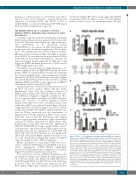
The analysis was performed by taking advantage of co- culture systems of Scr or J1/2KD HMCL grown on a layer murine (NIH3T3) or human (HS5) stromal cells to measure the variations in SDF1α gene or protein expression. Results obtained by qRT-PCR with murine-specific primers (Figure 5A) indicate that HMCL promoted the activation of Notch signaling (HES5) and SDF1α gene expression in NIH3T3 cells, which could be reverted by J1/2KD.
Similar results were observed at protein level as assessed by flow cytometry analysis (Figure 5B and Online Supplementary Figure S10) on co-cultures composed of HMCL and the human GFP+ HS5 cells. Of note, the selec- tive inhibition of Jagged1 or Jagged2 is clearly less effec- tive if compared with the simultaneous J1/2KD, that max- imizes the outcome on SDF1α inhibition (Online Supplementary Figure S11). Flow cytometric results were validated by ELISA on conditioned media (Figure 5C) indi- cating that MM cell-derived Jagged can increase SDF1α production by BMSC. We further confirmed that the vari- ation in SDF1α expression was the consequence of Jagged-activated Notch signaling in BMSC by an assess- ment that showed that the stimulation with Jagged1 and/or Jagged2 peptides can increase HS5 cell-mediated secretion of SDF1α, measured by ELISA (Figure 5D). Additionally, we knocked down Notch1 expression in HS5 cells (N1KD HS5) by using a specific siRNA, as previ- ously reported,11 and observed that SDF1α expression sig- nificantly decreased in comparison to control HS5 cells (Figure 5E). Since Notch1 silencing does not significantly affect HS5 cell viability (Online Supplementary Figure S12), we could exclude the possibility that reduction of SDF1α expression might be due to HS5 cell apoptosis.
administration administration administration
On the other hand, we verified that J1/2KD was associ- ated to a reduced CXCR4 expression in HMCL used in co- culture experiments. J1/2KD HMCL significantly decreased CXCR4 expression in comparison to Scr HMCL (Figure 5F and Online Supplementary Figure S13).
Figure 3. Effect of J1/2 inhibition on multiple myeloma (MM) cells ability to promote bone marrow (BM)-induced drug resistance. (A) A Notch-responsive dual luciferase assay was carried out in HS5 cells cultured alone or in the pres- ence of Scr or J1/2KD human multiple myeloma cell lines (HMCL) for 24 hours (h). Data were normalized on luciferase activity in HS5 cells cultured alone (=100). Mean±standard deviation of three independent experiments are shown. Statistical analysis was performed using one-way ANOVA and Tukey post-test (*P<0.05; **P<0.01; ***P<0.001). (B and C) Co-cultures of J1/2KD or Scr HMCL with the BM stromal cell (BMSC) line HS5 were established to evaluate the effect of J1/2KD on BMSC-induced drug resistance. The experi- mental timeline is reported. Graphs show the percentage of apoptotic OPM2 (B) or U266 (C) cells (Annexin V+/GFP–). Values of apoptosis of each type of cul- ture (Scr alone, Scr + HS5 and J1/2KD + HS5) treated with drugs are normal- ized to the corresponding controls treated with DMSO. Results are shown as mean±standard error of at least three independent experiments. Statistical analysis was performed using Kruskal-Wallis and Dunn post-test (*P<0.05; **P<0.01).
We assessed the outcome of SDF1α stimulation on the anti-apoptotic background of HMCL by analyzing the lev- els of Survivin, BCL2, and ABCC1 in U266 cells treated with 500 ng/mL SDF1α for 48 h. We observed an increase in Survivin, BCL2, and ABCC1 gene expression by qRT- PCR analysis (Figure 5G) confirmed at protein level by
B
C
haematologica | 2020; 105(7)
1929
