
Page 186 - 2020_07-Haematologica-web
P. 186
M. Colombo et al.
trend of protection of HMCL from apoptosis induced by Bor (15% in OPM2 and 26% in U266), Melph (20% in OPM2 and 11% in U266), and Len (14% in OPM2) (Figure 3B and C), although the statistical significance was reached only in the case of OPM2 treated with Bor. Conversely and more importantly, J1/2KD induced a statistically significant increase in apoptosis, re-establishing HMCL drug sensitiv- ity by hampering BMSC-mediated protection (HS5 cells do not display any significant increase in apoptosis; data not shown). Notably, although U266 cells were resistant to Len treatment in culture alone or in the presence of HS5 cells, apoptotic rate increased up to approximately 20% upon J1/2KD. The basal apoptotic effect of J1/2 KD on MM cells cultured with HS5 cells is reported in Online Supplementary Figure S6. As before, the selective Jagged1 or Jagged2 silenc- ing was less effective than the simultaneous J1/2KD (Online Supplementary Figure S7).
Since HS5 cells could act as a source of paracrine/autocrine Jagged ligands, we wondered why they cannot rescue J1/2KD in MM cells. Western blot analysis indicates that the expression levels of Jagged1 and
Jagged2 in HS5 cells are significantly lower than those expressed by OPM2 and U266 cells (Online Supplementary Figure S8). This can reasonably explain why, in our co-cul- ture system, Notch signaling activated in HMCL by BMSC is not sufficient to rescue the loss of Jagged1 and Jagged2 in MM cells.
We further explored whether Jagged-mediated Notch activation in BMSC could promote the pharmacological resistance of MM cells by up-regulating the anti-apoptotic effectors previously analyzed, Survivin, BCL2, and ABCC1. To evaluate gene expression changes, we took advantage of a co-culture system including OPM2 or U266 cells with a non-human mimic model of BMSC, the murine cell line of NIH3T3 fibroblasts. This approach enabled us to precisely assess the expression levels of human (HMCL-derived) anti-apoptotic genes in co-culture by using species-specific primers. Results showed that BMSC were able to promote the expression of the anti- apoptotic effectors Survivin, BCL2, and ABCC1 in Scr HMCL, while BMSC co-cultured with J1/2KD HMCL lost this ability (Figure 4A and B). Importantly, using an entire-
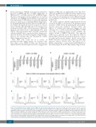
B vs.
A
vs.
C
D
Figure 2. J1/2 withdrawal affects multiple myeloma (MM) cells anti-apoptotic background. We analyzed how J1/2KD affects Notch activation and the expression of anti-apoptotic genes in human multiple myeloma cell lines (HMCL). (A and B) Confirmation of J1/2KD efficacy in OPM2 (A) and U266 (B) cells was obtained by quantitative polymerase chain reaction (qPCR) assay assessing the relative gene expression variation of Jagged1 and Jagged2 and Notch target genes HES1 and HES6 (normalized to GAPDH) in cells transfected with J1/2 siRNA compared to cells transfected with Scr siRNA, calculated by the 2-ΔΔCt formula. The expression levels of the anti-apoptotic effectors BCL2, Survivin, and ABCC1 were also analyzed. Data are expressed as mean± standard deviation of at least three independent exper- iments. Two-tailed t-test confirmed statistically significant downregulation of the tested genes; *P<0.05; **P<0.01; ***P<0.001. (C and D) Histograms display the levels of BCL2, Survivin and ABCC1 protein (black lines) analyzed by flow cytometry in J1/2KD OPM2 or Scr OPM2 (C) and J1/2KD U266 or Scr U266 (D) and an isotype-matched control (gray line). Histograms are representative of at least three independent experiments.
1928
haematologica | 2020; 105(7)
