
Page 120 - 2020_07-Haematologica-web
P. 120
J.N. Fisher et al.
A
B
C
DE
FG
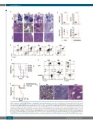
Figure 3. Expression of iNUP98-KMT2A induces a transplantable acute myeloid leukemia in some mice. (A) Histological sections and peripheral blood (PB) smears from four leukemic iNUP98-KMT2A mice. Scale bar: 10 μm. (A) Blood values from doxycycline (DOX)-treated iNUP98-KMT2A mice which developed symptoms of leukemia as well as wildtype (WT) littermate controls (CTRL). *P<0.05, **P<0.01, unpaired t-test, n=5. WBC: white blood cells; LUC: abnormal leukocytes; RBC: red blood cells. (C) Gr-1 and Mac-1 expression on total BM cells from leukemic iNUP98-KMT2A mice and a representative control mouse. (D) Total BM from leukemic mice M1 and M2 was transplanted into WT recipients in the presence or absence of DOX. Kaplan-Meier curves show disease-free survival of transplanted animals. **P<0.01, log-rank test. (E) Representative immunophenotypes of total BM of recipients (on DOX) (from Figure 3D) either transplanted with iNUP98-KMT2A acute myeloid leukemia from mouse M1: M1a and M1b; or from mouse M2. (F) iNUP98-KMT2A mice were exposed to DOX 48 h prior to sublethal irradiation (600 cGy). Irradiated (IR) WT mice were used as controls. Kaplan-Meier curves illustrate disease-free survival. *P<0.05, log-rank test. (G) Representative histopathology section of a symptomatic irradiated iNUP98-KMT2A mouse. The BM is infiltrated with blasts which are also visible in the peripheral blood (PB) and kidney. Scale bars: PB: 10 μm; BM & kidney: 100 μm.
1862
haematologica | 2020; 105(7)
