
Page 73 - Haematologica - Vol. 105 n. 6 - June 2020
P. 73
Innate drug responses in hematologic cell populations
(Online Supplementary Table S1) in 96-well plates to pro- vide a direct comparison between cell types derived from healthy donors and those derived from patients with iden- tical immunophenotypes. Moreover, this enabled detec- tion of drug responses in rare cell subsets, such as plasma cells (CD138+) and progenitor cells (CD34+CD38- or CD34+CD38+).
While ex vivo response to the proteasome inhibitor bortezomib was detected in most cell types (Figure 2B), CD138+CD38- plasma cells were resistant compared to CD138+CD38+ or other cells (Online Supplementary Figure S5). A higher response to the nucleoside analog clofara- bine was noted for CD3+CD4- and CD34+CD38+ cells compared to CD3+CD4+ or CD34+CD38- cells. Dexamethasone depleted CD19+ and CD56+ cells and induced a dose-dependent increase in the CD14+ cell count (Figure 2B). T-cell subsets were insensitive to PI3K/mTOR inhibitor omipalisib. A similar effect for sev- eral molecules targeting the PI3K-mTOR signaling axis was observed in CD3+ enriched cells tested with a cell via- bility assay (Online Supplementary Figure S6). Surprisingly, an increase in CD3+ cell count was noted at concentrations of 10 and 100 nM (Figure 2B). Apart from individual vari- ations, distinct drug efficacies associated with healthy cell
A
lineages were detected equally in all patient specimens (Figure 2B and Online Supplementary Figure S7).
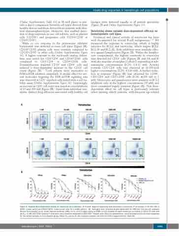
Venetoclax shows variable dose-dependent efficacy on hematopoietic cell types
Preclinical and clinical activity of venetoclax has been well documented for several B-cell malignancies.17-20 We measured the response to venetoclax, which is highly selective for BCL2, and navitoclax, which targets BCL2, BCL-W and BCL-XL. Both inhibitors were similarly effec- tive against lymphocytes (Figure 2B). Within the lympho- cyte compartment, the highest sensitivity to venetoclax was detected for CD19+ cells (Figures 2B, and 3A and B) with the majority of samples (Cohort I) responding at sub- nanomolar concentrations (IC50, 0.4-12 nM). Activity towards CD3+CD4- cells was observed at 10-100-fold higher concentrations (IC50, 8-140 nM). A further reduc- tion in response (Figure 3B) was observed for CD56+, CD3+CD4+ and CD3+CD56+ cells (IC50, ≅100 nM to 1 μM). Monocytes and granulocytes were sensitive to BCL2 inhibitors only at the highest concentration (10 μM) and were considered largely resistant (Figure 2B). This dose- dependent effect on cell types is particularly relevant when treating elderly patients, with frequent age-related
B
Figure 3. Variable dose-dependent activity of venetoclax on leukocytes. (A) Scatter diagram displaying dose-dependent cytotoxicity of venetoclax (1-10,000 nM) in CD45+ (upper panel) and CD45+CD19+ (lower panel) cells for a single patient. (B) Averaged dose response graphs generated for different immune cell subtypes derived from healthy (n=3), acute myeloid leukemia (AML) (n=3), and multiple myeloma (MM) (n=10) samples showed venetoclax sensitivity in CD19+/B cells with an IC50 <1 nM. CD3+CD4- cytotoxic T cells were more sensitive compared to CD3+CD4+ T-helper cells. Data are presented as mean±standard error of mean responses for the tested samples in each disease group. Mean IC50 values for the analyzed samples are listed in Online Supplementary Table S4.
haematologica | 2020; 105(6)
1531
