
Page 283 - Haematologica - Vol. 105 n. 6 - June 2020
P. 283
Platelet depletion for effective transfusion
remaining platelet population during DT treatment (Figure 1E). Notably, linear regression of platelet counts until day 4 revealed that their decrease corresponded to their reported lifespan (Figure 1F) of 4.8 days.10 Platelets were therefore not directly affected by DT treatment and thrombocytopenia was induced by megakaryocyte deple- tion rather than platelet ablation. Examining a putative immune response against DT, we quantified DT-specific immunoglobulin G (IgG) generated during treatment. Antibodies were detectable from day 8 onwards, with no significant difference between iDTRPlt and WT mice, indi- cating that presence of iDTR on platelets did not alter the adaptive immune response (Figure 1G). Furthermore, leukocyte count remained unaffected in thrombocy- topenic iDTRPlt mice (Figure 1H). Upon prolonged platelet depletion mice displayed an increased burden of hematomata and impaired wound healing, with body weight being slightly, but not significantly decreased (Figure 1I). We also observed that male mice fared worse than females (data not shown). Therefore, only female mice were used in further experiments.
Presence of iDTR does not affect platelet function
To monitor potential off-target effects of iDTR expres- sion on platelet physiology, we performed blood counts as well as platelet function assays. Untreated iDTRPlt mice
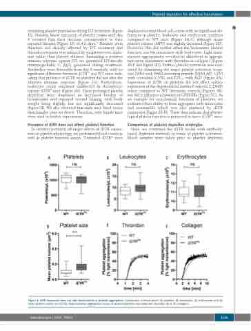
displayed normal blood cell counts with no significant dif- ferences in platelet, leukocyte and erythrocyte numbers compared to WT mice (Figure 2A-C) although mean platelet volume (MPV) was slightly increased (Figure 2D). However, this did neither affect the hemostatic platelet function, nor the interaction with leukocytes. Light trans- mission aggregometry revealed no alterations in aggrega- tion upon stimulation with thrombin or collagen I (Figure 2E-F and Figure 3D). Further, platelet activation was eval- uated by stimulating the major platelet activation recep- tors PAR4 with PAR4-activating peptide (PAR4-AP), GPVI with convulxin (CVX), and P2Y1/12 with ADP (Figure 3A). Expression of iDTR on platelets did not affect surface expression of the degranulation marker P-selectin (CD62P) when compared to WT littermate controls (Figures 3B), nor did it influence activation of GPIIb/IIIa (Figure 3C). As an example for non-classical functions of platelets, we evaluated their ability to form aggregates with monocytes and neutrophils which was also unaltered by iDTR expression (Figure 3E-H). These data indicate that physio- logical platelet function is preserved in naive iDTRPlt mice.
Comparison of platelet depletion strategies
Next, we compared the iDTR model with antibody- based depletion methods in terms of platelet activation. Blood samples were taken prior to platelet depletion
ABC
DEF
Figure 2. iDTR expression does not alter blood counts or platelet aggregation. Comparison of blood count: (A) platelets, (B) leukocytes, (C) erythrocytes and (D) mean platelet volume (n=13-14). Representative aggregation curves of washed platelets stimulated with thrombin (E) or (F) collagen I.
haematologica | 2020; 105(6)
1741
