Page 210 - Haematologica - Vol. 105 n. 6 - June 2020
P. 210
  I. Scheller et al.
 Critical determinants of thrombus formation are the locally prevailing rheological conditions. With increasing shear rate, e.g. during the development of stenosis, platelet adhesion and aggregate formation become increasingly dependent on GPIb-vWF interactions. At sites of very high, pathological shear rates and disturbed flow, occlusive arterial thrombus formation can be mediated predominantly by the GPIb-vWF interaction, does not involve visible platelet shape change or activation, and may thus be mediated, at least in part, by the biomechan- ical interaction of platelets.2 In accordance with this, it was shown that vWF-mediated pulling at the GPIbα receptor under shear induces the unfolding of a juxtamembrane mechanosensitive domain, which might contribute to platelet mechanosensing under dynamic conditions and GPIb-induced intracellular signaling.3 Thus, it appears that platelet-mediated thrombus formation in vivo involves both agonist receptor and biomechanical signaling, the interplay of which is still poorly understood.
Platelets are produced by megakaryocytes (MK) in the bone marrow (BM) through a cytoskeleton-driven process. The critical role of the actin cytoskeleton for platelet production and function is illustrated by the asso- ciation of mutations in genes encoding proteins that are involved in actin cytoskeletal organization, such as Diaphanous Related Formin 1 (DIAPH1),4 filamin A (FLNA),5 Wiskott Aldrich syndrom protein (WASP),6 actinin 1 (ACTN1),7 or tropomyosin 4 (TPM4)8 with platelet disorders in humans and mice. In circulating platelets, the actin cytoskeleton is essential to maintain cell morphology and to exert key functions upon activa- tion, such as granule release, as well as the formation of filopodia and lamellipodia.9 However, the complex protein network orchestrating actin dynamics in platelets is not fully understood.
The ADF homology domain (ADF-H) is one of the best- characterized actin binding motifs. The ADF-H protein family comprises twinfilin (Twf), ADF/n-cofilin, Abp1/drebrin, and coactosin-like 1 (Cotl1/CLP). We have previously shown that Twf2a and ADF/n-cofilin play dis- tinct, critical roles in platelet formation and function.10,11 Although sharing less than 20% sequence identity with the other ADF-H family members, Cotl1 is structurally highly homologous, suggesting a similar role for cytoskeletal dynamics.12 Indeed, Cotl1 binds F-actin, but does not interact with actin monomers.13 Furthermore, Cotl1 was shown to prevent n-cofilin-mediated depoly- merization of actin filaments, thereby promoting lamel- lipodia formation at the immune synapse.14 Besides its interaction with F-actin, Cotl1 is implicated in the biosyn- thesis of leukotrienes (LT),15 lipid-derived pro-inflamma- tory mediators involved in a variety of inflammatory processes such as allergy or asthma. Cotl1 was shown to interact with 5-lipoxygenase (5-LO), a key enzyme in LT biosynthesis that catalyzes two of the initial steps, namely the oxygenation of arachidonic acid (AA) to 5-HPETE and the subsequent dehydration into the epoxide LTA4.15-17 Platelet-stored LT have been shown to contribute to inflammatory responses, e.g. during lung inflammation.18 However, the mechanism underlying this contribution, as well as the precise role of LT for platelet function, have not been defined.
Here, we generated conditional knockout mice lacking Cotl1 in the MK lineage. We found that Cotl1 is critically required for stable platelet thrombus formation under con-
ditions of shear flow in vitro and in vivo by modulating the function of the mechanoreceptor GPIb, as well as platelet LT biosynthesis.
Methods
Animals
Cotl1+/- mice were generated by injection of embryonic stem cell clone Cotl1tm1a(EUCOMM)Hmgu into C57Bl/6 blastocysts. Germline trans- mission was confirmed by backcrossing of the chimeric mice with C57Bl/6 mice. Cotl1+/- mice were intercrossed with mice carrying Flp recombinase to generate Cotl1fl/+ mice, which were intercrossed with mice carrying Cre recombinase under control of the platelet factor 4 (Pf4) promoter to generate mice lacking Cotl1 specifically in MK and platelets.19 For all experiments, 12- to 16-week old Cotl1fl/fl;Pf4Cre and Cotl1fl/fl littermate controls, maintained on a C57Bl/6 background, were used. All mice were derived from the following breeding strategy: Cotl1fl/fl;Pf4Cre X Cotl1fl/fl..
Animalstudies
Animal studies were approved by the district government of Lower Franconia (AZ15_14; Bezirksregierung Unterfranken).
Further details of reagents and experimental procedures are given in the Online Supplementary Appendix.
Results
Cotl1 deficiency does not affect platelet function under static conditions
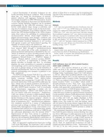
Cotl1fl/fl;Pf4Cre mice (hereon referred to as Cotl1-l-) were viable and born in the expected Mendelian ratios. The absence of Cotl1 in platelets was confirmed by western blotting (Online Supplementary Figure S1A). Cotl1 deficien- cy did not affect either peripheral platelet count, size or ultrastructure (Figure 1A-C) nor the expression of promi- nent platelet surface receptors (Online Supplementary Table S1). This is in contrast to deficiency in the ADF-H protein family members Twf2a11 or n-cofilin,10 which results in thrombocytopenia in mice. Thus, Cotl1-/- mice represent the first knockout mouse model of an ADF-H family member with apparently normal platelet biogenesis.
A common feature of ADF-H family members is their involvement in cytoskeletal dynamics. Consistently, n- cofilin deficiency resulted in impaired stimulus-dependent F-actin assembly, whereas Twf2a-deficient mice displayed increased actin dynamics.10,11 According to previous stud- ies, n-cofilin and Cotl1 are highly abundant in both human and mouse platelets,20,21 while Twf2 levels are slightly lower. The results for Twf1 and drebrin, which was not listed in the mouse dataset, suggest that they are expressed at a lower level. Importantly, both datasets indi- cate that the expression of ADF-H proteins is similar in human and mouse platelets.
We determined expression levels of the ADF-H mem- bers Twf1/2a and n-cofilin by western blotting, but could not detect differences in total expression levels of either protein between WT and Cotl1-/- platelets (Figure 1D and E). Strikingly, we observed a strong (2-fold) increase in expression of the (inactive) phosphorylated form of n- cofilin (Ser3) in Cotl1-/- platelets (***P<0.001) (Figure 1E and F). Next, we analyzed the localization of Cotl1 in resting and spread platelets (Online Supplementary Figure S1B and C). In line with previous observations,22 we
   1668
haematologica | 2020; 105(6)


