
Page 143 - Haematologica - Vol. 105 n. 6 - June 2020
P. 143
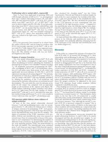
 Follicular helper T cells in FL
   Proliferating cells in contact with TFH express AID
Since TFH have been implicated in initiating SHM and CSR through induction of AID in GC,20 we investigated if there was a spatial relationship between TFH and AIDpos cells. AID was restricted to Ki67pos cells in FL, 63% ±8.8 of which were AIDpos and 39.8% ±9.7 of AIDposKi67pos cells were in direct contact with PD-1Hi cells. As we had estab- lished that most PD-1Hi cells in contact with Ki67pos cells were ICOSpos, we can predict that the majority of PD-1Hi cells in contact with AIDposKi67pos cells were TFH (Online Supplementary Figure S5). AID was similarly restricted to Ki67pos cells of GC where close association with PD-1Hi cells was evident in the light zones (Online Supplementary Figure S5C).
PD-L1
PD-L1 has previously been reported to be absent from the surface of FL B-cells36 and we found no evidence that PD-L1 was strongly expressed on the Ki67pos cells in con- tact with PD-1Hi cells. Instead, PD-L1 was expressed main- ly on interfollicular CD23neg cells (Online Supplementary Figure S6). The identity of these cells was not further investigated in this study.
Features of synapse formation
The close spatial relationship between Ki67pos FL B cells and TFH was further investigated in high power images where morphological features indicating the formation of immunological synapses were identified. Features included: TFH cell membrane projections encompassing the Ki67pos cells, overlapping of the B- and T-cell membranes, distor- tion of T-cell nuclei away from points of cell contact and significantly increased intensity of CD4, PD-1, and ICOS expression at points of cell contact (Figure 5).37 The intensity of expression of CD4, PD-1, and ICOS at points where TFH were in contact with Ki67pos cells was formally quantified by defining the perimeter of the T-cell membrane and measuring the intensity of fluorescence at each pixel around the perimeter. The area under the curve for intensity at the point of cell contact was compared with an equivalent length of cell membrane at the opposite pole (Figure 5B). Similar features indicating synapse formation were identi- fied in GC light zones but the high number of closely- packed Ki67pos cells and TFH in GC precluded the same ana- lytic method being used because TFH in GC were usually in contact with more than one Ki67pos cell simultaneously.
TCR repertoire within follicles shows evidence of antigen restriction
In view of the close spatial relationship observed between T cells and proliferating tumor cells, we investi- gated whether T cells within the follicles show evidence of antigen restriction by performing TCRV next genera- tion sequencing of genomic DNA from laser dissected fol- licular and interfollicular areas from five FL samples. The degree of restriction of the TCRV repertoires in FL neo- plastic follicles and interfollicular areas was assessed in several ways. First, we estimated the richness of the reper- toire in each compartment by determining the number of different clones present per ng of input DNA which, since we were analysing genomic DNA, was proportionate to the total cell number. The interfollicular areas contained more T-cell clones per ng of input DNA than the intrafol- licular regions, however, this did not quite reach statistical significance (P=0.06, Online Supplementary Table S4). We
also calculated the clonality index38 (see the Online Supplementary Materials and Methods for further details). In each of the five cases examined, the clonality of the follic- ular T cells was greater than in the interfollicular areas (P=0.0317, Figure 6A). We also calculated the proportion of the TCR repertoire in each compartment that was accounted for by high frequency clones.39 Compared to the interfollicular areas, the follicular regions were domi- nated by high frequency clones (Figure 6B). For example, the top 50 most frequent clones made up a mean of 19% of all clones in the follicular areas (95% CI: 17-21) com- pared to 9.8% in the interfollicular region (95% CI: 6.1- 13.4) P=0.0002, n=5.
As expected from their different phenotypes, the clones present in the follicular and interfollicular areas of the same sample were markedly different, indicating that the TCR repertoires of the follicular and interfollicular areas are distinct (Figure 6C).
Discussion
In this study we compared the structure of neoplastic fol- licles in FL with GC in reactive LN, focusing on TFH, their relationship with proliferating B cells and TCR repertoire. Although TFH have previously been reported to be present in the FL microenvironment,24-27 these studies were per- formed on disaggregated LN and this is the first time that their spatial organization has been investigated in situ in this way. Using multi-parameter immunofluorescent confo- cal microscopy, we demonstrated that TFH – as identified by surface expression of CD4, PD1, and ICOS - constitute a similar proportion of CD4pos T cells in FL as in reactive LN and form synapses with proliferating Ki67pos tumor cells which express the DNA modifying enzyme AID. The num- ber of TFH in neoplastic follicles correlates with the level of tumor proliferation and histological grade, and there is evi- dence for antigen restriction, as supported by the more clonal TCR repertoire found within neoplastic follicles compared to interfollicular areas.
These findings are novel and of significance for a number of reasons. First, in contrast to previous work on disaggre- gated FL LN, which showed an increase in the total number of TFH ,24,25,40 we found that FL follicles contain TFH in similar proportions to normal reactive GC. This discrepancy likely relates to the fact that in FL, LN architecture is usually effaced by many closely packed follicles, whereas in normal tissues the interfollicular areas, which contain many fewer TFH, are more extensive. Thus, although the overall TFH con- tent of FL LN is increased compared to normal,25 this is because of the larger number of follicles in the tumor and when neoplastic and normal follicles are compared directly, the numbers are the same. This finding underlines the need to complement data obtained from disaggregated tissues with anatomic studies.
Our use of multiparameter microscopy permitted the spatial relationship between TFH and B cells to be closely investigated and this also provided new insights. TFH are essential for providing normal GC B cells with signals nec- essary for their survival, proliferation and maturation.19,41 To our knowledge, this is the first time that the intimate rela- tionship between Ki67pos B cells and TFH has been demon- strated in situ in human LN in this way and our observa- tions are in keeping with the pivotal role they play in the normal GC reaction. Importantly, we also found that the
haematologica | 2020; 105(6)
  1601
