
Page 270 - Haematologica May 2020
P. 270
J. Köhler et al.
mouse (C57BL/6)-derived plasma clots.21 To test whether S. pyogenes can survive in, and escape from, plasma clots of BALB/c mice, plasma samples from control- or PPK- ASO treated animals were clotted with thrombin in the presence of S. pyogenes. Clots were covered with 1% plas- ma and incubated for up to 4 h at 37°C. Viable S. pyogenes count assays from the supernatants and the clots were per- formed. Two hours after incubation, no bacteria could be detected in the supernatant. Four hours after incubation, the supernatants of PPK-depleted clots contained signifi- cantly fewer viable bacteria compared to supernatants from control-ASO clots (Figure 6A). Thus, S. pyogenes is able to escape from BALB/c mouse plasma clots and PK plays an important role in this process. The relative impor- tance of PK was confirmed in PPK-depleted plasma recon- stituted with human PK, where significantly more viable bacteria (compared to PPK-depleted plasma clots) could be detected in the supernatants (Figure 6A). No difference in CFU could be determined inside the clots (Figure 6B).
It has previously been shown that PK activates plas- minogen,22 which would enhance plasmin concentration in control mice. To address this experimentally, circulating plasmin 2-antiplasmin (PAP) complexes were quantified in infected control- and PPK-ASO treated mice (Figure 6C). However, no difference between the two groups was observed, supporting the assumption that PK assist in fib- rinogen degradation and might be relevant for the observed bacterial dissemination. Importantly, there was no difference in the plasminogen concentration between healthy control and PPK-ASO treated mice (Figure 6D).
Finally, plasma clots from control ASO or PPK-depleted animals incubated with S. pyogenes were imaged with
scanning electron microscopy. Clots derived from control mice and incubated with bacteria lost their structural integrity (Figure 6E), fibrin fibers were degraded, and bac- teria could not be detected within the clot (Figure 6E, left). If PPK was absent, intact fibrin fibers were visible, with bacteria trapped inside of them (Figure 6E, right).
Taken together, the data show that PK supports degra- dation and streptococcal escape from mouse plasma clots leading to increased bacteremia and infection of tissues.
Plasma kallikrein and FXII concentrations are significantly decreased in plasma of septic patients
We further investigated PK and FXII concentrations in plasma of 23 patients with sepsis, severe sepsis or septic shock, collected 1, 2 and 3 days after admission to the Intensive Care Unit (ICU). In most of the patients, multi- ple infectious sources and bacterial isolates were identified and, importantly, non-survivors had significantly pro- longed aPTT compared to survivors.14 Plasma samples from 12 healthy persons were used as controls. Both PK and FXII levels are significantly reduced in all patients compared to healthy controls (Figure 7A and B). There is no significant difference between survivors and non-sur- vivors, or between the days of admission. The data sup- port earlier studies, and clearly show that reduction of PK and FXII in plasma is not restricted to S. pyogenes sepsis.
Discussion
Local activation of contact factors on the bacterial sur- face may be protective against infections, due to the
AB
C
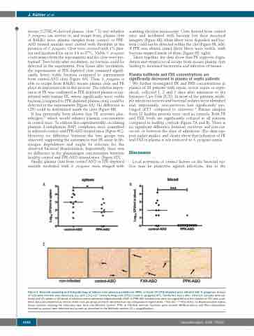
Figure 3. Bacterial spreading and histopathology of kidneys from plasma prekallikrein (PPK)- or factor XII (FXII)-depleted mice infected with S. pyogenes. Groups of mice were infected subcutaneously (sc.) with 1.6-2 x10⁷ colony forming units (CFU)/mouse S. pyogenes AP1. Twenty-four hours after infection, samples were col- lected and (A) spleen or (B) blood of infected control-antisense-oligonucleotide (ASO) or PPK-ASO treated mice were homogenized and the number of CFU was quan- tified. Data are presented as means of ten mice per group and were obtained from two independent experiments. *P≤0.05; ***P≤0.0001. (C) Representative kidney tissue sections showing the medullary rays, from non-infected, control-, PPK, or FXII-ASO animals. Sections were stained (MSB-Lendrum) and fibrin depositions (marked by arrows) were detected and scored as described in the Methods section (10 x magnification).
1430
haematologica | 2020; 105(5)
