
Page 206 - Haematologica May 2020
P. 206
L. Chen et al.
chemotaxis.10 Therefore, molecular depletion or chemical inhibition of SYK or pan-PI3K blockade may limit CXCR4 internalization and increase residual cell surface CXCR4 expression. However, SYK/PI3K inhibition also increases nuclear localization of FOXO1 and associated FOXO1- mediated transactivation of CXCR43,8,13,30 For these rea- sons, we depleted FOXO1 in a BCR-dependent DLBCL cell line (DHL4), treated the cells with vehicle or R406 and subsequently measured CXCR4 expression by flow cytometry (Online Supplementary Figure S3). SYK inhibition induced less CXCR4 in FOXO1-depleted cells (Online Supplementary Figure S3), highlighting the role of FOXO1 in CXCR4 expression.
A
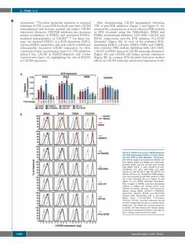
After demonstrating CXCR4 upregulation following SYK or pan-PI3K inhibition (Figure 1 and Figure 3), we assessed the consequences of more selective PI3K isoform or BTK blockade using the PI3Kα/d>β/γ, PI3Kd and PI3Kd/γ predominant inhibitors, GDC-0941, CAL101 and IPI145, respectively, and the BTK inhibitor, PCI-32765 (ibrutinib)9 (Figure 4A). In each of the evaluated BCR- dependent DLBCL cell lines (DHL4, DHL6 and TMD8), more selective PI3K isoform inhibition with GDC-0941, CAL101 or IPI145 increased CXCR4 transcript abundance (Figure 4A) and CXCR4 cell surface protein expression (Figure 4B). In contrast, BTK blockade had more modest effects on CXCR4 transcript and protein expression in the
B
Figure 4. CXCR4 expression in BCR-dependent and BCR-independent DLBCL cell lines follow- ing SYK, PI3K or BTK inhibition. BCR-depen- dent diffuse large B-cell lymphoma (DLBCL) cell lines (DHL4, DHL6 and TMD8) and a BCR-inde- pendent DLBCL cell line (Toledo) were treated with DMSO, R406 (1 mM), GS-9973 (2 mM), LY294002 (10 mM), GDC-0941 (0.5 mM), CAL101 (2 mM), IPI145 (1 mM), PCI-32765 (0.1 μM) for 24 hours (h). Thereafter, CXCR4 expres- sion was analyzed by qRT-PCR relative to PPIA (A) or flow cytometry (B) as in Figure 1A and Figure 1C. (A) CXCR4 transcript abundance. Fold changes in CXCR4 transcript abundance relative to DMSO are shown below each inhibitor for the four cell lines. The P-values for vehicle versus R406, GS-9973, LY294002, GDC-0941, CAL101, IPI145 and PCI-32765 treated were determined with a one-sided Welch t-test. ***P<0.0001; **P<0.001; *P<0.01; #P<0.05. Error bars represent the SD of three independent assays in a representative experiment. (B) CXCR4 cell surface expression in DLBCL cell lines treated with vehicle (black) or the above-mentioned inhibitors (see key) for 24 h. Isotype-matched control in gray.
1366
haematologica | 2020; 105(5)
