
Page 164 - Haematologica May 2020
P. 164
V. Agnusdei et al.
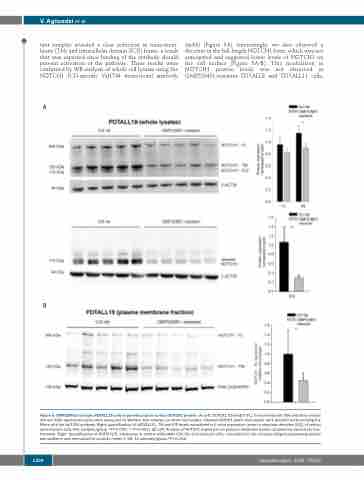
tant samples revealed a clear reduction in trans-mem- brane (TM) and intracellular domain (ICD) forms, a result that was expected since binding of the antibody should prevent activation of the pathway. These results were confirmed by WB analysis of whole cell lysates using the NOTCH1 ICD-specific Val1744 monoclonal antibody
A
(mAb) (Figure 5A). Interestingly, we also observed a decrease in the full-length NOTCH1 form, which was not anticipated and suggested lower levels of NOTCH1 on the cell surface (Figure 5A-B). This modulation in NOTCH1 protein levels was not observed in OMP52M51-resistant PDTALL8 and PDTALL11 cells,
B
Figure 5. OMP52M51-resistant PDTALL19 cells show reduction of surface NOTCH1 protein. (A) Left: NOTCH1 full-length (FL), transmembrane (TM) and intra-cellular domain (ICD) expression levels were measured by Western blot analysis on whole cell lysates. Cleaved NOTCH1 levels (mid panel) were determined by probing the filters with the Val1744 antibody. Right: quantification of NOTCH1 FL, TM and ICD levels normalized to β-actin expression (mean ± standard deviation [SD]) of control and resistant cells (five samples/group. *P=0.005; **P<0.001). (B) Left: Analysis of NOTCH1 expression on plasma membrane lysates obtained by subcellular frac- tionation. Right: Quantification of NOTCH1-FL expression in control antibodies (Ctrl Ab) and resistant cells, normalized to the corresponding housekeeping protein pan-cadherin and normalized to controls (mean ± SD, 10 samples/group.*P=0.001).
1324
haematologica | 2020; 105(5)
