
Page 80 - Haematologica April 2020
P. 80
L.A. Hampton O’Neil et al.
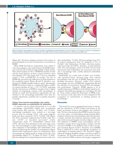
Figure 6. Schematic representation of the role of receptors in erythroblastic island development. Summary diagram of the receptors involved in macrophage-ery- throblast binding in erythroblastic island development in the VCAM+ cells of the bone marrow compared to ex vivo culture and VCAM– cells.
(Figure 4D). Therefore, integrin activation at the surface of proerythroblasts is not due to the presence or activation of EPHB4.
When EPHB knockdown erythroblasts were added to +Dex macrophages, only erythroblasts with EPHB4 deple- tion caused a loss of both average number of links (scram- bled control median of 0.43 vs. EPHB4 KD median of 0.26) and the mean duration of these contacts between +Dex macrophages (75% last longer than 1 hour in scrambled control vs. 50% in EPHB4 KD on average). The loss of EPHB6 did not lead to any differences in mean duration to the scrambled control (Figure 4E and F); therefore, con- firming the EPHB4 peptide inhibitor result. Interestingly, EPHB6 knockdown leads to a statistically higher number of contacts (median of 0.43 vs. 0.57; P<0.0001), indicating that the sole presence of EPHB4 is enough to have a higher number of long-lasting contacts with macrophages. It should be noted that knockdown experiments are less sensitive to the macrophage:erythroblast ratio than the peptide experiments, probably due to peptide saturation occurring.
Primary bone marrow macrophages also require EPHB4 expression on erythroblasts for interaction
To extend a role for ephrin interactions to ex vivo BM macrophages, erythroblastic islands were reconstituted using human BM aspirates. VCAM1+ and VCAM1- macrophage populations were isolated in a two-stage process, first by isolating CD14+ cells using magnetic beads and then sorting for CD14+ VCAM1+ cells (Figure 5A). Flow cytometry confirmed that VCAM1+ cells were also CD169+ as described previously39 (Online Supplementary Figure S1). Erythroblasts cultured from CD34+ cells were then introduced at the same stage as for the +Dex macrophages in earlier experiments. Clusters of cells composed of macrophages and erythroblasts were observed following this method of reconstitution (Figure 5B and Online Supplementary Figure S5). Furthermore, these macrophages are highly motile as witnessed for the +Dex macrophages (Figure 5C).40 In Figure 5D and E, VCAM1+ BM macrophages have long-lasting interactions with mul-
tiple erythroblasts. VCAM1- BM macrophages have 50% of contacts lasting more than 1h compared to 75% of VCAM1+ cells. Furthermore, VCAM1- cells form statisti- cally fewer links (P<0.0001). Therefore, as demonstrated with +Dex macrophages, a lack of VCAM1 does not stop interactions with erythroblasts, but its presence does indi- cate a macrophage with a subtly enhanced erythroblast binding ability.
Importantly, the results with VCAM1+ and VCAM1– cells reproduced those observed using +Dex cultured macrophages with the loss of EPHB4 impacting on ery- throblast association. However, unlike +Dex cultured macrophages, the loss of EPHB6 also significantly affected the initiation of associations between BM macrophages and erythroblasts. Therefore, EPHB6 appears to be as equally important as EPHB4 in erythroblastic island recon- stitutions when BM macrophages are used (Figure 5D and E). These results confirm the importance of EPHB recep- tors’ presence at the same time as active integrins for the recognition of erythroblasts as binding partners by macrophages.
Discussion
This work has used an imaging-based assay to interro- gate the importance of EPH receptor interactions in the initial association between macrophages and erythrob- lasts. We have demonstrated that EPHB receptors are pres- ent at the surface of erythroblasts during terminal differ- entiation between the proerythroblast and orthochromat- ic stages, and that this temporal expression profile coin- cides with increased integrin activation (Figures 1 and 2). The depletion of EPHB4 causes macrophages and ery- throblasts to have fewer long-duration contacts, and the removal of EPHB4 or its inhibition had no effect on inte- grin activation (Figures 4 and 5). Therefore, loss of EPHB4 alone is sufficient to reduce macrophage recognition of erythroblasts as binding partners. We, therefore, suggest ephrin-B2 binding alongside integrin activation reinforces recognition. We therefore propose a new model of inter- action whereby EPHB receptors and integrin engagement
922
haematologica | 2020; 105(4)
