
Page 283 - Haematologica April 2020
P. 283
c.2269_2270del vWF and non-canonical splicing site
were undetectable. rvWF multimer analysis revealed larg- er than normal oligomers, and a delayed oligomer migra- tion (Figure 5).
SDS-PAGE and western blot analysis - Analyzing samples with SDS-PAGE and western blotting in reducing condi- tions revealed a protein band with a molecular weight cor- responding to the unprocessed vWF-pp (about 340KDa) in both P2 and P3 rvWF, while a band corresponding to the mature cleaved vWF (250KDa) was seen in the WT. These data confirmed the persistence of vWFpp in the P2 and P3 vWF (Figure 6).
Discussion
We report on a complex splicing alteration associated with the vWF c.2269_2270del mutation that involves the
use of a non-canonical splicing site in a case of severe type 1 vWD characterized by a lack of multimer organization in the circulating vWF. The c.2269_2270del mutation was described in 2003 as a pure gene null mutation.29 Here, we demonstrate that it alters vWF splicing, and induces the generation of three different aberrant mRNA species. The predominant specie, RNAI, encodes for the already- known truncated p.Leu757Valfs*22 vWF (P1 in the text), associated with a pure quantitative vWF defect.29 The other two species found, RNAII and RNAIII, account for less than 20% of the total vWF mRNA expression, and are produced by the activation of two cryptic acceptor splic- ing sites (a canonical “AG” and an unusual “CG”) in exon 18 and intron 17 of the vWF gene, respectively. RNAII encodes for a mature vWF characterized by loss of the furin cleavage site (p.L757_R763delinsVSSQ, P2), while RNAIII has an altered furin recognition site that impairs its
Table 1. Main hemostatic and genetic findings in the proband and his parents.
Patients
Proband
Mother Father Son Daughter
Normal range
aPTT PFA100 RIPA FVIII:C VWF:Ag VWF:CB VWF:CB VWF:FVIIIB VWF:FVIIIB PlateletVWF BS
sec
54.6
22.8 31.0 97.9 93.2
24-36
sec % U/dL U/dL U/dL
>300 0(5.6)* 3.4 1.2 1.9
160 72.9 209.6 114.3 104.9
198 74.2 89.1 39.3 40.4
150 65.4 119.6 66.0 61.4
145 72.3 108.2 56.3 63.6
94-193 72±12 60-160 60-160 65-150
ratio U/dL ratio U/dL
1.58 0 NC 1.7 30
0.91 118.0 1.03
1.03 38.2 0.97
0.93 61.1 0.97
1.13 55.0 0.97
≥0.75 65-150 ≥0.74
97.6 1 103.0 0 74.4 1 83.9 1
70-140 0-3/0-5 (M/F)
aPTT: activated partial thromboplastin time; RIPA: ristocetin induced platelet aggregation at 1.2 mg/mL ristocetin concentration; *RIPA value at 1.5 mg/mL ristocetin concentra- tion; BS: Bleeding Score, calculated by the ISTH Bleeding Assessment tool (BAT); M/F: male/female; NC: not calculable.
A
B
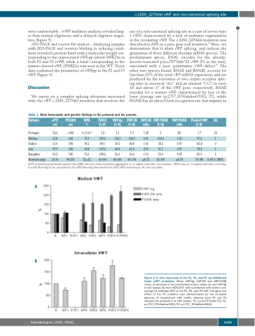
Figure 4. In vitro expression of the P1, P2, and P3 von Willebrand factor (vWF) mutations. Mean vWF:Ag, vWF:CB and vWF:FVIIIB ratios, as obtained in the conditioned culture media (A), and vWF:Ag in cell lysates (B) from HEK293T cells transfected with vectors con- taining the wild-type (WT) or the P1, P2, and P3 vWF. The gene null effect of the P1 mutation was demonstrated by the complete absence of recombinant vWF (rvWF), whereas both P2 and P3 induced the production of vWF protein. P1: p.Leu757Valfs*22; P2: p.L757_R763delinsVSSQ; P3: p.L757_761delinsVSSQG.
haematologica | 2020; 105(4)
1125
