
Page 285 - Haematologica April 2020
P. 285
c.2269_2270del vWF and non-canonical splicing site
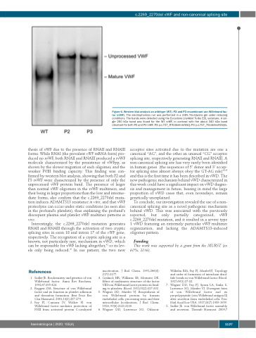
thesis of vWF due to the presence of RNAII and RNAIII forms. While RNAI (the prevalent vWF mRNA form) pro- duced no rvWF, both RNAII and RNAIII produced a rvWF molecule characterized by the persistence of vWFpp, as shown by the slower migration of each oligomer, and the weaker FVIII binding capacity. This finding was con- firmed by western blot analysis, showing that both P2 and P3 rvWF were characterized by the presence of only the unprocessed vWF protein band. The presence of larger than normal vWF oligomers in the rvWF multimers, and their being in larger proportions than the low and interme- diate forms, also confirm that the c.2269_2270del muta- tion induces ADAMTS13 resistance in vitro, and that vWF proteolysis can occur under static conditions (as seen also in the proband’s platelets), thus explaining the proband’s discrepant plasma and platelet vWF multimer patterns in vivo.
Interestingly, the c.2269_2270del mutation generates RNAII and RNAIII through the activation of two cryptic splicing sites in exon 18 and intron 17 of the vWF gene, respectively. The recognition of a cryptic splicing site is a known, not particularly rare, mechanism in vWD, which can be responsible for vWF lacking altogether,42 or its lev- els only being reduced.43 In our patient, the two new
acceptor sites activated due to the mutation are one a canonical “AG”, and the other an unusual “CG” acceptor splicing site, respectively generating RNAII and RNAIII. A non-canonical splicing site has very rarely been identified in human genes (the sequences of 5’ donor and 3’ accep- tor splicing sites almost always obey the GT-AG rule)44,45 and this is the first time it has been described in vWD. The new pathogenic mechanism behind vWD characterized in this work could have a significant impact on vWD diagno- sis and management in future, bearing in mind the large proportion of vWD cases that, even nowadays, remain genetically unexplained.
To conclude, our investigation revealed the use of a non- canonical splicing site as a novel pathogenic mechanism behind vWD. This was associated with the previously reported, but only partially categorized, vWF c.2269_2270del mutation, and it resulted in a severe type 1 vWD featuring an extremely particular vWF multimer organization, and lacking the ADAMTS13-induced oligomer pattern.
Funding
This work was supported by a grant from the MURST (ex 60%, 2016).
References
1. Sadler JE. Biochemistry and genetics of von Willebrand factor. Annu Rev Biochem. 1998;67:395-424.
2. Ruggeri ZM. Structure of von Willebrand factor and its function in platelet adhesion and thrombus formation. Best Pract Res Clin Haematol. 2001;14(2):257-279.
3. Fay PJ, Cumans JV, Walker FJ. von Willebrand factor mediates protection of FVIII from activated protein C-catalyzed
inactivation. J Biol Chem. 1991;266(4):
2172-2177.
4. Gralnick HR, Williams SB, Morisato DK.
Effect of multimeric structure of the factor VIII/von Willebrand factor protein on bind- ing to platelets. Blood. 1981;58(2):387-392.
5. Wagner DD, Marder VJ. Biosynthesis of von Willebrand protein by human endothelial cells: processing steps and their intracellular localization. J Biol Chem. 1984; 99(6):2123-2130.
6. Wagner DD, Lawrence SO, Ohlsson-
Wilhelm BM, Fay PJ, MarderVJ. Topology and order of formation of interchain disul- fide bonds in von Willebrand factor. Blood. 1987;69(1):27-32.
7. Wagner DD, Fay PJ, Sporn LA, Sinha S, Lawrence SO, Marder VJ. Divergent fates of von Willebrand factor and its propolypeptide (von Willebrand antigen II) after secretion from endothelial cells. Proc Natl Acad Scie USA. 1987;84(7):1955-1959.
8. Sadler JE. von Willebrand factor assembly and secretion. Thromb Haemost. 2009;7
Figure 6. Western blot analysis on wild-type (WT), P2 and P3 recombinant von Willebrand fac- tor (rvWF). The electrophoresis run was performed in a 3-8% Tris-Glycine gel under reducing conditions. The bands were detected using the Euroclone LiteAblot Turbo ECL substrate. A sin- gle 250 kDa band was found for the WT rvWF, in contrast with the about 340 kDa band observed for both P2 and P3 rvWF. P2: p.L757_R763delinsVSSQ; P3: p.L757_761delinsVSSQG.
haematologica | 2020; 105(4)
1127
