
Page 269 - Haematologica April 2020
P. 269
Inflammation and TTP
AB
C
DEF
G
HIJ
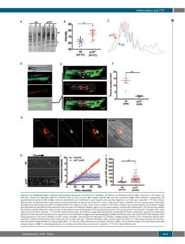
Figure 2. von Willebrand factor multimer and thrombus formation in wt and a13-/- zebrafish. (A) Plasma von Willebrand factor (VWF) multimers in wt (lanes 1-3) and a13-/- (lanes 4-6) zebrafish aged 3-4 months. Here, H and L on the right margin indicate high and low molecular weight VWF multimers, respectively. (B) Quantification of plasma VWF antigen levels by densitometry (all multimers in each sample were scanned together) in wt and a13-/- zebrafish. **P<0.01, Mann- Whitney test. (C) Representative densitometric scanning profiles of plasma vwf multimers in a13-/- (red) and wt (blue) zebrafish. (D) The caudal areas of zebrafish endogenously expressing fli-1/eGFP and gata-1/sRed: the square or ovals show where a drop of 0.3% ferric chloride was placed topically. (E) Confocal images demonstrated the presence of thrombocytes (red) and loss of endothelial integrity (green) in the caudal vessels (outlined with dotted lines). The red arrows indicate the direction of blood flow from artery to vein. (F) Quantitative data showing the time to complete occlusion in the caudal vessel in wt and a13-/- zebrafish after injury induced by 0.3% ferric chloride. ***P<0.005, Mann-Whitney test. (G) Confocal image demonstrated the incorporation of both erythrocytes (red) and thrombocytes (green) into the occlusive thrombus in the caudal vein of wt zebrafish endogenously expressing gata-1/dsRed (red blood cells) and cd41/eGFP (thrombocytes) after being exposed to 0.3% ferric chloride. (H) The surface coverage of fluorescent thrombocytes on a fibrillar collagen-coated surface in the microfluidic channel after perfusion of diluted whole blood (1:20), obtained from wt (top) and a13-/- (bottom) zebrafish, under arterial shear (15 dyne/cm2). Red arrows indicate the thrombo- cyte-decorated strings formed on a vwf/collagen fiber. (I, J) The rate of fluorescence accumulation (means ± SEM, n=3) (I) and the lengths of thrombocyte-decorated strings (means ± SEM) (J), following perfusion of a diluted whole blood from wt and a13-/- zebrafish. **P<0.01, Mann-Whitney test.
haematologica | 2020; 105(4)
1111
