
Page 24 - Haematologica April 2020
P. 24
C. Côme et al.
continued from the previous page
2019
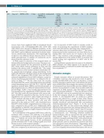
Song et al.34
MISTRG or NSG
1-3 days
yes, 2x150cGy
(MISTRG) or 1x100cGy (NSG)
intrahepatically
1 del(5q),
3 MLD,
2 RS-SLD,
1 RS-MLD,
1 MPN-RS-T, 3 EB-1, 3 EB-2 1 MLD, 2 EB-1, 1 EB-2
BM CD34+
BM CD3-
0.2-2.75x10⁵
1.15-6x10⁵
No 22
No 4
12-31 weeks
13-15 weeks
The main characteristics of the protocols published to generate patient-derived xenograft (PDX) models from patients with myelodysplastic syndromes (MDS) are presented. NOD/SCID: non- obesediabeticmicewithPrkdcscid mutation;β2m-/-:beta2-microglobulinnull;3/GM/SF:constitutiveexpressionofhumaninterleukin-3(IL-3),granulocyte-macrophagecolony-stimulatingfactor (GM-CSF) and stem cell factor (SF); NOG: NOD/Shi-scid Il2rgnull; NSG: NOD.Cg- Prkdcscid Il2rgnull;; NSG-S: NSG with human IL-3, GM-CSF and SF constitutive expression; MISTRG: humanized macrophage colony-stimulating factor, IL-3/GM-CSF, SIRP alpha and thrombopoietin combined with Rag2-/-, IL2Rγ-/-; i.v.: intravenous injection; i.p: intraperitoneal injection; cGy: centigray; BM: bone marrow; PB: peripheral blood; MNC: mononuclear cells; MSC: mesenchymal stromal cells; HSC: hematopoietic stem cells; Lin-: lineage negative; RA: refractory anemia; RAEB: refractory anemia with excess blasts; RAEB-I: RAEB type 1; RAEB-II: RAEB type 2; RAEBt: RAEB in transformation; RARS: refractory anemia with ringed sideroblasts; RARS-T: RARS with thrombocytosis; RS: ringed sideroblasts; RCMD: refractory cytopenia with multilineage dysplasia; RCMD-RS: RCMD and ringed sideroblasts; del(5q): MDS associated with isolated deletion of chromosome 5q; MDS/MPN: myelodysplastic/myeloproliferative neoplasms; CMML: chronic myelomonocytic leukemia; JMML: juvenile myelomonocytic leukemia; MLD: multilineage dysplasia; SLD: single lineage dysplasia; EB: excess blasts; MDS/MPN-RS-T: MDS/MPN with ring sideroblasts and thrombocytosis; n.s.: not specified.
sources have been employed (BM or peripheral blood mononuclear cells, CD3-depleted BM cells, CD34+ BM cells) which were injected in different quantities, in the presence or absence of BM-derived mesenchymal stromal cells (MSC) and in different anatomical locations (intra- venous, intrafemoral, intrahepatic). Not surprisingly, this resulted in very different disease latencies (from 3 to 32 weeks post-injection). A number of conclusions can be drawn from this extensive work:
(i) With respect to selection of the recipient strain, an immunodeficient background is necessary. The most com- monly used recipient for the generation of PDX is the NSG strain which harbors mutations in Prkdc and Il2g leading to the absence of B, T and NK cells.35 The consti- tutive expression of the human cytokines interleukin-3 (Il- 3), granulocyte-macrophage colony-stimulating factor (GM-CSF) and stem cell factor (SCF) on this background (NSG-S, also designated NSG-SGM3) does not lead to enhanced engraftment of most MDS subtypes, except for chronic myelomonocytic leukemia,28 in contrast to the sit- uation in AML.24,26,29,36 On the other hand, the recently developed MISTRG strain, expressing human macrophage colony-stimulating factor (M-CSF), IL-3, GM-CSF, signal regulatory protein alpha (SIRPα) and thrombopoietin at physiological levels on a different immunodeficient back- ground (Rag2-/-, IL2Rγ-/-), was recently demonstrated to be a promising host for engraftment of MDS patients’ mate- rial.34 Not only could cells from patients with various sub- types of MDS be expanded in this line, but the levels of engraftment were increased, with a higher percentage of CD33+ myeloid cells than in NSG mice. Moreover, long- term engraftment of these myeloid cells was also improved in this strain as CD33+ cells constituted more than 80% of the hCD45+ compartment in secondary recip- ients, compared to 30% in NSG mice. Additionally, MDS cells engrafted in MISTRG mice generated erythroid and megakaryocytic lineages at a higher frequency than in the NSG counterpart.34
(ii) T-cell depletion of the primary MDS tumor, either by treatment with a human CD3 antibody or by physical separation, is a prerequisite to limit graft-versus-host dis- ease.26,34,36-38 Indeed, one of the first attempts to generate MDS PDX failed mainly because of the predominant growth of human CD3+ T cells, leading to graft-versus-host disease in most of the recipient animals.37
(iii) Intrafemoral injections result in better engraftment in NSG mice compared to an intravenous route of injection.
(iv) Co-injection of MSC leads to variable results in terms of promoting the engraftment of MDS samples, with some laboratories reporting some enhancement,22-24 whereas others have not found this effect.26,29 The under- lying reasons for this variation are not clear. However, as human MSC only survive for 2-4 weeks in the murine BM,24,26 this variation could potentially reflect patient- specific differences in the ability of MSC to promote the initial seeding and engraftment of MDS cells in the murine BM.
(v) Engraftment capacity does not seem to be related to MDS subtypes, but rather appears to be specific to the individual samples, as indicated in studies with large num- bers of patients.24,26,34
Alternative strategies
Despite extensive efforts in several laboratories, this cumulative work has only produced a total of approxi- mately 100 MDS PDX so far. There is, therefore, a strong need for alternative systems that could enhance the gener- ation of MDS PDX. Interestingly, descriptions of a number of humanized bone marrow-like structure (hBMLS) mod- els have been published recently. These models enable the expansion of AML patients’ cells that failed to engraft with conventional methods.39-41 They are all based on the use of BM MSC and can be separated into two categories. In the first category, which we will define as “scaffold” models, in vitro-expanded MSC are seeded in a gelatin sponge and cultured for a couple of days. Next, human leukemic cells are injected into the sponge which is subse- quently introduced subcutaneously into non-irradiated immunocompromised mice41 (Figure 1). In the second approach, BM MSC are first mixed with Matrigel and introduced subcutaneously into immune-deficient mice in which they develop a so-called “ossicle” after 2-3 months, which constitutes an exterior bone structure surrounding a hematopoietic core. Following sublethal irradiation, human leukemic cells are injected into the ossicle where they expand39 (Figure 1). Another ossicle-like approach combines osteogenic priming of MSC with a physical sup- port consisting of two or three biphasic calcium phos- phate particles, prior to subcutaneous insertion into mice and subsequent ossicle development.40 Importantly, up to four hBMLS per animal can be introduced,39-41 and Reinisch et al. have elegantly demonstrated that tumor cells can cir-
866
haematologica | 2020; 105(4)
