
Page 208 - Haematologica April 2020
P. 208
V. Griggio et al.
BAY87-2243 restores fludarabine sensitivity of TP53dis chronic lymphocytic leukemia cells and counteracts the protective effect of stromal cells
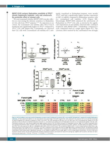
We next examined whether BAY87-2243 was also effec- tive in overcoming the intrinsic resistance to fludarabine of CLL cells from TP53dis patients.9,31-33 As expected, in our cohort, the normalized cell viability after 48-h F-ara-A treatment was significantly higher in TP53dis compared to TP53wt CLL cells (Figure 6A). Consistently, we observed that CLL cells with a normalized cell viability ≥0.5, arbi-
trarily considered as fludarabine-resistant, were mostly TP53dis and had a significantly higher baseline expression of HIF-1A mRNA compared to fludarabine-sensitive cells (i.e. normalized cell viability <0.5) (Figure 6B). Interestingly, BAY87-2243 enhanced the cytotoxicity of fludarabine on TP53dis CLL cells, as shown by the signifi- cant impairment of cell viability observed after combined treatment with BAY87-2243 + fludarabine compared to each compound used as a single agent (Figure 6C). The cytotoxic effect exerted by the combination was strongly
D
AB
C
Figure 6. HIF-1A mRNA is over-expressed in fludarabine-resistant chronic lymphocytic leukemia (CLL) cells and HIF-1α inhibition is capable of restoring fludarabine sensitivity. (A) Normalized cell viability (i.e. the ratio between the percentage of AnnV/PI negative CLL cells cultured in the presence of F-ara-A and the percentage of AnnV/PI negative CLL cells left untreated) of TP53dis and TP53wt CLL cells exposed for 48 hours (h) to 10 μM F-ara-A. (B) A normalized cell viability of 0.5 was selected as the cut-off value to identify fludarabine-resistant (i.e. normalized cell viability ≥0.5) and fludarabine-sensitive (i.e. normalized cell viability <0.5) CLL cells. Fludarabine-resistant CLL cells showed significantly higher baseline levels of HIF-1A mRNA compared to fludarabine-sensitive. Eight of 10 (80%) fludarabine-resis- tant samples derived from TP53dis patients, and 13 of 14 (93%) fludarabine-sensitive samples derived from TP53wt patients. (C) Normalized cell viability of TP53dis and TP53wt CLL cells exposed for 48 h to 1 μM BAY87-2243 (BAY) and/or 10 μM F-ara-A. The combination BAY87-2243 + F-ara-A (striped pattern) determined a sig- nificant decrease in the viability of TP53wt and TP53dis CLL cells, compared to each compound used as single agent and to untreated controls. (D) Heatmaps showing normalized viability after 48-h treatment with BAY87-2243 + fludarabine as single agents or in combination, used at different concentrations. Asterisks indicate the combinations which determined a significant reduction in cell viability compared to each single agent, at the corresponding concentration. (A and C) Box plots represent median values and 25-75% percentiles; whiskers represent minimum and maximum values for each group, together with all points. (B) Data are repre- sented as bee-swarm plot. *P<0.05; **P<0.01; ***P<0.001; ****P<0.0001.
1050
haematologica | 2020; 105(4)
