
Page 318 - Haematologica March 2020
P. 318
P. Ferraresi et al.
c.64+196A, c.131-394C, c.292-672A, c.571+78A, M1 (Haplotype 2). The analysis was also extended to the c.571+78G>A heterozygotes, which revealed that haplo- type 1 is compatible with patients of Maghreb origin whereas haplotype 2 is compatible with patients of European origin.
The c.572-392C>G, c.64+305G>A and c.806-329A vari- ants were also relatively frequent as they were found in three, two and two alleles, respectively, whereas the remaining were identified only once.
Computational analysis of splicing regulatory elements
We performed an in silico analysis of the six deep- intronic mutations to infer a pathogenic effect on splicing. In principle the nucleotide changes could affect Intronic Splicing regulatory elements such as enhancers (ISE) or Silencers (ISS)43 or create/strengthen 5’ or 3’ splice sites (ss). However, regulatory elements generally reside with- in the first 200 bp of the intron44 and the main bioinfor- matics tools (i.e. Human Splicing Finder, www.umd.be/HSF/) have been developed to predict exonic elements, which confers prediction of the impact of the investigated changes with an unacceptable degree of speculation. Therefore, we focused the analysis on 5’ss and 3’ss (www.fruitfly.org/seq_tools/splice.html), which pre- dicted that the c.64+305G>A, c.291+846C>T, c.681+132G>T nucleotide changes do not appreciably strengthen cryptic splice sites. Concerning the c.571+78G>A, c.572-392C>G and c.806-329G>A vari- ants, the introduction of the nucleotide changes would result in the creation (c.572-392C>G) or remarkable strengthening (c.571+78G>A and c.806-329G>A) of a cryptic 5’ss (Table 2).
In vitro characterization of the splicing variants
Based on the bioinformatics prediction of the impact of these variants on splicing, on the number of affected alle-
les, on the MAF, and on identification in homozygous conditions, we selected the c.571+78G>A, c.572-392C>G and c.806-329G>A changes for further characterization. Due to the impossibility of investigating F7 mRNA pro- cessing in patients’ hepatocytes, the physiological site of F7 synthesis, or to the unavailability of fresh leukocytes as ectopic source of F7 mRNA, we exploited the expres- sion of F7 minigenes (Figure 2A). The transfection of the pIVS6-wt minigene and splicing pattern analysis revealed correct splicing (Figure 2C, transcript 2) but also, albeit to a lesser extent, exon 6 skipping (transcript 1) and usage of the weak cryptic intronic 5’ss at position +79 that leads to partial intron retention (transcript 3G).
The splicing pattern analysis of cells transfected with the pIVS6+78A minigene showed an aberrant transcript (Figure 2C, transcript 3A) that, upon sequencing, indicat- ed the usage of the strengthened intronic 5’ss at position +79 (Figure 2B, transcript 3A). This leads to partial intron retention resulting in a deleted and frame-shifted mRNA harboring a premature nonsense triplet at position p.201, not expected to produce a functional FVII protein. To evaluate the presence of residual FVII levels, the RT-PCR was fluorescently labeled and the amplicons evaluated by denaturing capillary electrophoresis, which ensures high sensitivity. This approach led us to identify very low levels of correct transcripts, which roughly account- ed for approximately 3% of the overall transcripts (Figure3B).
While the splicing analysis of c.806-329G>A construct in pCDNA3 did not reveal any alteration by transient transfection in HEK293T (Online Supplementary Appendix and Online Supplementary Figure S1) or in Baby Hamster Kidney cells (data not shown), the assessment of splicing pattern of cells transfected with the FVII minigene har- boring the c.572-392C>G change revealed splicing abnor- malities (Figure 2B). In addition to the correctly spliced mRNA, we identified transcripts arising from skipping of
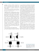
Figure 1. Family pedigree of patient #28. The propositus is indicated by an arrow. Mutation, nucleotide changes, A1A2 (rs5742910), M1M2 (p.Arg413Gln, rs6046) haplotypes, coagulant factor VII (FVII:C) and factor VII anti- gen (FVII:Ag) are indicated.
832
haematologica | 2020; 105(3)
