
Page 320 - Haematologica March 2020
P. 320
P. Ferraresi et al.
Discussion
Uncharacterized F7 pathogenic alleles are mentioned in all patient databases, and NGS would represent a power- ful tool to tackle this; however, so far, this has not been fully explored. Here, we applied this approach in 13 FVII deficient patients who were only partially characterized through conventional sequencing, and identified a panel of deep intronic substitutions as candidates to explain the reduced FVII:C levels in patients. However, as for the numerous deep intronic nucleotide changes identified by NGS and associated with inherited diseases, their patho- genic role requires experimental support.
The evidence for the pathogenicity of deep intronic variations relies on several clinical and molecular observa- tions. The first aspect to consider is their virtual absence in databases. This led us to exclude from our selection the c.64+305G>A and c.291+846C>T variants, with a minor allelic frequency of 0.005 and 0.004, respectively. The same applies to the c.681+132G>T change, reported in dbSnp databases as rs752129277 but with an estimated frequency of <0.0004. Another aspect that could suggest the pathogenicity of the nucleotide changes is their distri- bution among affected relatives or non-related individu-
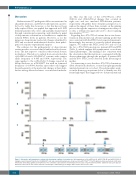
A
B
C
als. This was the case of the c.571+78G>A, c.572- 392C>G and c.806-329G>A changes that occurred in eight, two and two unrelated FVII-deficient patients, respectively. Altogether these elements prompted us to explore the impact of these three variants on the splicing process through the expression of minigenes in eukaryot- ic cells, a well-proven approach used to dissect splicing abnormalities.4,13,14,42,45
Regarding the c.571+78G>A variant, the in vitro charac- terization demonstrated an aberrant splicing profile that was consistent with the FVII:C levels reported in patients. Interestingly, the amount of correctly spliced transcripts (approx. 3%) reflects the FVII:C levels (3% and <1%) in the two c.571+78A homozygotes (patients #15 and #90) (Table 1), which explains the asymptomatic or moderate clinical phenotypes. This finding is also consistent with the observation that the mutation co-segregated with the disease phenotype through the pedigree of patient #28, and the 50% FVII:C levels detected in the heterozygous mother.
It is interesting to note that the c.571+78G>A mutation, albeit absent from databases, occurred in eight apparently unrelated patients in our cohort. The polymorphic analy- sis in the homozygous patients led us to identify two dif- ferent haplotypes that suggested two distinct mutational
Figure 3. Alternative splicing patterns evaluated by denaturing capillary electrophoresis. Splicing patterns of the pIVS6-wt (A), pIVS6+78A (B) and pIVS6-392G (C) minigenes upon transient transfection in HEK293T cells and evaluated by polymerase chain reaction with T7bisFFam-F7ex7R oligonucleotides performed at 28 cycles followed by denaturing capillary electrophoresis. The schematic representation of transcripts (not in scale) is reported below and the amplicons base pairs (bp) are indicated at the bottom. The relative amount of transcripts is indicated by percentages. RFU: relative fluorescence units.
834
haematologica | 2020; 105(3)
