
Page 317 - Haematologica March 2020
P. 317
NGS of F7 and minigene studies identify molecular bases of FVII deficiency
pipelines: SEQUENCE Pilot® module SeqNext, version 4.3.1 (JSI medical systems GmbH, Ettenhein, Germany) and SeqOne® (https://app.seq.one). The dataset had a mean coverage of over 300x. For each nucleotide of interest, the sequence depth was at least of 30x and the Phred-based quality score above Q30. Regions that did not reach these criteria were sequenced using Sanger technology (primers available upon request); this also applies to each deleterious variation that was independently checked using Sanger sequencing.
Expression studies with F7 minigenes
The expression vectors for the c.571+78G>A (pIVS6+78A)
and c.572-392C>G (pIVS6-392G) variants were created by site- directed mutagenesis of the pIVS6 wild-type (pIVS6-wt), created by cloning the F7 genomic region spanning exon 5 through exon 7 into the pcDNA3 plasmid.4 Changes were introduced, as pre- viously described,40 through the overlapping oligonucleotides 5’GAAGCAGATCAAAAGTAAGCATGGGATC3’ and 5’GATCC- CATGCTTACTTTTGATCTGCTTC3’ for the c.572-392C>G mutation. For the c.571+78G>A mutation, we exploited the non-overlapping oligonucleotides 5’CTGGACAAAA- GACAGGTGGG AGTGGC3’ and 5’TAAGATAATCCG- TAGTGGGACAGGG ACT3’ in a slightly modified protocols that implies, after PCR cycles, the addition of T4 polynucleotide kinase and T4 DNA ligase to ensure circulation of the products.
The U7smOPT expression vectors (pU7smOPT) were created as previously described41 using a standard SP6 reverse oligonu- cleotide 5’ATTTAGGTGACACTATAG3’ and the forward muta- genic oligonucleotides 5’ACAGAGGCCTTTCCGC AcccacctgtcttttgtccaAATTTTTGGAG3 ’ (U7+78A), 5’ACAGAGGCCTTTCCGCAtgaagccactcccacctgAATTTTTGG AG3’ (U7+78Ash) and 5’ACAGAGGCCTTTCCGCAcatgcttact tttgatctAATTTTTGGAG3’(U7-392G).
One microgram of the pIVS6 variant, alone or with equimolar amounts of the pU7smOPT variant, was transiently transfected in human embryonic kidney cells (HEK293T) by lipofection in 12-well plates.13 RNA isolation and reverse transcription42 were followed by PCR using the forward oligonucleotide T7bisF (5’CACTGCTTACTGGCTTATCGAAAT3’, in the pcDNA3 T7 region), either unmodified or 5’fluorescently labeled (T7bisFFAM), and the reverse oligonucleotide F7ex7R (5’CACAACTGAGCTC- CATTCACCAACA3’, in exon 7) or F7PsExR (5’TTCAATCAAG- GTCTTGGGCC3’, in the pseudo-exon 5b).
Results
Genotyping of FVII deficient patients
Among the 13 selected FVII-deficient patients shown in Table 1, ten had FVII:C levels below 15% and were
expected to have two F7 pathogenic alleles. However, the conventional sequencing did not reveal any F7 pathogen- ic allele for patients #15, #90 and #330, and only one for the remaining seven patients (#17, #19, #28, #31, #113, #214, #262). On the other hand, three patients (#284, #341, #377) presented with FVII:C levels between 15% and 30% but displayed only the c.430+1G>A mutation (#341) or the A2M2 polymorphic haplotype (#284, #377), which points towards the presence of an additional F7 pathogenic allele for each patient. In this scenario, we had a total of 16 F7 uncharacterized alleles to be explored using NGS.
As far as the clinical phenotype is concerned, 3 of 13 patients were symptomatic. Patient #19 presented with bruises and frequent epistaxis, patient #90 had post-trau- matic oral bleeding, spontaneous hematuria and rectal bleeding, and patient #214 suffered from provoked hematoma and severe menorrhagia resolved by replace- ment therapy.
Next-generation sequencing, besides confirming the presence of the causative variants identified by the Sanger approach, also revealed several deep intronic substitu- tions. Among them, only those with a coverage of 30x and observed in databases with a minor allele frequency (MAF) <0.05 were analyzed further. Six deep intronic substitutions matched these criteria (Table 2). The c.571+78G>A change, whose pathogenic effect is sup- ported by its co-segregation with the disease phenotype in the family pedigree of patient #28 (Figure 1), was the most frequent in our series, being present in ten alleles from unrelated patients living in various areas, including France, North Africa and Lebanon (Table 1). NGS data prompted us to analyze an enlarged panel of polymorphic deep intronic variants in both c.571+78G>A homozy- gotes (#15, #90), who were homozygous for the major A1 and M1 polymorphic alleles. However, they differed on other intronic variants. Patient #15, of Tunisian origin, was homozygous for two variants, c.-402A (rs510317) and c.292-672G (rs12431329), that are quite rare, with a minor allele frequency (MAF) of 0.233 and 0.213, respec- tively. By contrast, patient #90, living in Lebanon, showed the c.-402G and the c.292-672A variants, and displayed two additional deep intronic polymorphic variants in the homozygous state, the c.64+196G>A (rs2774030) and the c.131-394T>C (rs1745939), with a global frequency of the c.64+196A and c.131-394C alleles of 0.551 and 0.739, respectively. Thus, two different haplotypes associated with the c.571+78G>A mutation could be defined: c.- 402A, A1 c.64+196G, c.131-394T, c.292-672G, c.571+78A, M1 (Haplotype 1) and c.-402G, A1,
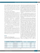
Table 2. Deep intronic mutations found by next-generation sequencing screening.
Change Intron
c.64+305G>A 1a
c.291+846C>T 2 c.571+78G>A 5 c.572-392C>G 5 c.681+132G>T 6
c.806-329G>A 7
Prediction
Weakening cryptic 3’ss
Strengthening cryptic 3’ss
Strengthening cryptic 5’ss
Creation of new 5’ss
Strengthening cryptic 3’ss
Strengthening cryptic 5’ss
Score (wt/mutated)
0.31/0.13
0.51/0.71
0.11/0.79
nd/0.98
0.02/0.12
0.79/0.99
Position rs MAF
-8 36208414 0.005
-7 565185989 0.004
-2 764741909 none
+1 none none
-8 752129277 none
+3 none none
Position of the point mutation is referred to the 5' splice site (5’ss) or 3’ss. RS: reference SNP ID number; MAF: minor allele frequency, based on 1000Genome project (http://www.internationalgenome.org). F7 gene reference sequence is NG_009262.1. NNSPLICE 0.9 software (www.fruitfly.org/seq_tools/splice.html) was used to predict and calculate the 5’ss or 3’ss of the score. Introns are indicated by legacy nomenclature. wt: wild-type.
haematologica | 2020; 105(3)
831
