
Page 105 - Haematologica March 2020
P. 105
Role of PIEZO1 during human erythropoiesis
Figures S8 and S9. Cultures of cells from four patients could be driven beyond day 10 and showed progressive terminal maturation and enucleation, but at a heteroge- neous rate, confirming that erythroid differentiation was delayed but not totally blocked (Online Supplementary Figure S10).
Discussion
The mechanotransductor PIEZO1 has a well-described role in regulating hydration and volume of mature ery- throcytes.12,28 Indeed, activating mutations are responsible for most HX cases and, recently, a frequent polymorphism (E756del) has been associated with resistance to malaria in African populations,29,30 although its influence on red cell hydration status is still controversial.31 PIEZO1 expression during erythropoiesis has been previously studied using RNA-sequencing analysis, the results of which were in agreement with our data that PIEZO1 is expressed in early progenitors14 and decreases during terminal maturation.23 These findings were confirmed at a protein level by exten- sive proteomic analyses of human erythroid differentia- tion.24 Whether PIEZO1 has a specific role during erythro- poiesis is not known, although evoked in recent case reports.19,32 Our data show for the first time that PIEZO1 is expressed and functional in erythroid progenitors, and that its activation influences erythroid differentiation, both in leukemic cell lines and primary erythroid cells. Indeed, PIEZO1 activation maintained cells at an imma- ture GPAlow stage for longer and tilted the transcriptional balance in favor of genes associated with an immature stage, such as GATA2 and BMI1, at the expense of genes associated with terminal differentiation, such as GATA1, GPA, ALAS2 or α and β globin without myeloid bias.33,34 Of
note, we used the chemical activator YODA1 to evaluate the effects of PIEZO1 on erythropoiesis.25 The phenotype was dependent on PIEZO1 since it was abrogated after Sh-RNA mediated PIEZO1 knockdown, and was also con- firmed in erythroid cells from HX patients carrying PIEZO1 gain-of-function mutations. Interestingly, we observed that PIEZO1 knockdown using a Sh-RNA strat- egy enhanced erythroid differentiation in UT7 cells. Since PIEZO1 activation was associated with an undifferentiat- ed state in UT7 cells, we may assume that a low PIEZO1 level at the cell surface could lead to a lower “basal” acti- vation rate and favor cell differentiation at the expense of cell proliferation. Of note, it was intriguing that a 50% reduction of PIEZO1 expression was sufficient to revert the YODA1-mediated effects. Although the GFP intensity determined the strength of YODA1 blockade, Sh-PIEZO1 significantly - but not totally - blocked the effects of YODA1 in GFP intermediate cells. This may argue for a threshold effect of PIEZO1, which could be further con- firmed in patch-clamp experiments.
PIEZO1 is known as a non-selective cation channel.7 We showed that the effect of PIEZO1 activation on erythro- poiesis was Ca2+-dependent, as described in mature red cells as well as in many other ‘PIEZO1-sensitive’ cells such as endothelial, urothelial, and epithelial cells.5,15–17 However, the consequences of a PIEZO1-mediated increase in intracellular Ca2+ may depend on the cell type. In mature red cells, the observed phenotype (i.e., red cell dehydration) occurs because of a secondary activation of the Gardos channel, which in turn exports K+ and induces loss of water.13 The effects of PIEZO1 on erythropoiesis seem not to occur through this pathway, since the Gardos inhibitor Senicapoc could not revert the phenotype. Ca2+ influx has previously been shown to be involved during erythropoiesis. Notably, it has been suggested that EPO
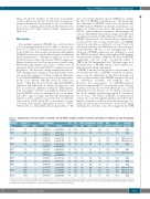
Table 1. Characteristics of the 14 patients, 11 families and 10 PIEZO1 mutations, number of patients and family per mutation and main hematologic features.
Mutation Patientn/ Id Familyn
HX#1 1/1
HX#2 2/2
PIEZO1mutation Age Exon cDNA Protein
14 c.1792G>A p.Val598Met 43
18 c.2344G>A p.Gly782Ser 55
18 c.2423G>A p.Arg808Gln
(mg/L) (mmol/g) experiments phenotype
Hb MCV Reticulocytes Ferritin MRI N.of Erythroid
42 c.6058G>A* p.Ala2020Thr 31
(g/L) (fL) x109/L
144 107 404
142 101 441
146 85 173 174 91 293
828 190
613 95
30 NA 980 104
N=3 High
N=3 High
N=1 Mild/ moderate N=1 Mild/ moderate
N=3 High
N=1 High
N=1 Mild/ moderate
N=1 High
N=1 High
N=2 High
N=1 Mild/ moderate N=1 Mild/ moderate N=1 Mild/ moderate
N=1 High
HX#3 3/3
4/3 42 c.6058G>A* p.Ala2020Thr 61
HX#4 5/4
HX#5 6/5
HX#6 7/6
HX#7 8/7
HX#8 9/8
HX#9 10/9
11/10
HX#10 12/10 13/10
HX#11 14/11
42 c.6007G>A p.Ala2003Thr 24
c.7471C>T p.Arg2491Trp
51 c.7391A>C p.His2464Pro 36
16 c.2152G>A p.Gly718Ser 51
c.7463G>A# p.Arg2488Gln
51 c.7529C>T p.Pro2510Leu 43 16 c.2042T>C p.Phe681Ser 44 51 c.7367G>A* p.Arg2456His 63
118 99 451 86 NA
181 87 205 71 NA
152 93 167 119 34
112 83 183 NA NA 145 102 248 86 15 112 120 288 535 200
16
c.2005G>T c.2005G>T c.2005G>T
p. Asp669Tyr p. Asp669Tyr p. Asp669Tyr
62 141 98 249 1000 330 39 142 104 496 889 NA 35 149 107 290 507 180
51 c.7367G>A* p.Arg2456His 78
129 85 221 600 NA
*Functional studies showing slower PIEZO1 inactivation kinetics.8,12 #: Functional studies showing a lower PIEZO1 activation threshold.8 Ferritin assays and magnetic resonance imaging evaluation of iron liver content were performed at diagnosis or before iron chelation/phlebotomy. Patients HX#9 and HX#11, from two different families, had the same PIEZO1 mutation. Id: identity, Hb: hemoglobin; MCV: mean corpuscular volume; MRI: magnetic resonance imaging.
haematologica | 2020; 105(3)
619
