
Page 160 - 2020_02-Haematologica-web
P. 160
A. Al Outa et al.
as compared to flies expressing the wild type variant BCR- ABL1p210 indicating that the transformation capacity of T315I is much higher than the wild type BCR-ABL1p210. Similar results were obtained when expressing BCR- ABL1p210/T315I in other tissues where more detrimental effects were seen when compared to BCR-ABL1p210. For example, expression of BCR-ABL1 in the fly imaginal discs resulted in pupal lethality with BCR-ABL1p210 expressing flies versus embryonic/larval lethality with BCR- ABL1p210/T315I expressing flies (unpublished data).
We further validated the model by assessing the capabil- ity of the conventional treatments used in clinics for CML patients of improving the eye defects observed in the adult eyes of BCR-ABL1p210 and BCR-ABL1p210/T315I flies. These TKI include imatinib as first generation TKI, nilo- tinib and dasatinib as second and ponatinib as third gener- ation TKI. Dasatinib and ponatinib resulted in the full res- cue of the BCR-ABL1p210 eye defect (Figures 5-6) in 100% and 86% of flies respectively. Imatinib and nilotinib (Figure 4; Online Supplementary Figure S1) exhibited a lower
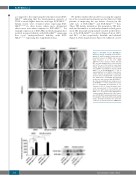
Figure 6. Ponatinib rescues BCR-ABL1p210 driven eye defect. Scanning electron micro- graphs of adult Drosophila compound eyes from flies fed on 0.3% DMSO only or pona- tinib. Posterior is to the left. GMR- GAL4>w1118 were used as control. E-H and M-P are high magnification of the posterior end of the eye in A-D and I-L respectively (692x). Normal development in control flies fed on DMSO or ponatinib is observed. BCR- ABL1p210 (C, G) and BCR-ABL1p210/T315I (K, O) expressing flies fed on DMSO show charac- teristic defective area with loss of ommatidi- al facets. Area is marked with a representa- tive dashed line. Ommatidial development in this area was restored with BCR-ABL1p210 flies fed on ponatinib (D, H). Compare to (C, G). BCR-ABLp210/T315I flies showed no restora- tion of ommatidial development (L, P). Compare to (K, O). Lower left panel repre- sents measurement of the posterior eye defect area (μm2). Data represents mean ± SEM. ****, P<0.0001. Lower right panel is a representative Western blot of the expres- sion of BCR-ABL1 and phosphorylated levels in transgenic untreated and treated adult fly heads. Genotypes indicated are under the control of eye specific promoter GMR-GAL4.
394
haematologica | 2020; 105(2)
