
Page 158 - 2020_02-Haematologica-web
P. 158
A. Al Outa et al.
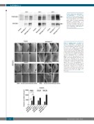
Figure 3. Expression of BCR-ABL1p210 and BCR-ABL1p210/T315I in the com- pound eyes. Representative Western blot of the expression of BCR-ABL1 and phosphorylated levels in trans- genic adult fly heads expressing BCR-ABL1p210 and BCR ABL1p210/T315I at different temperatures (18oC, 25oC, and 29oC). Genotypes indicated are under the control of eye specific pro- moter GMR-GAL4. GMR-GAL4>w1118 were used as control.
Figure 4. Imatinib shows a tendency to decrease BCR-ABL1p210 mediated eye defect. Scanning electron micrographs (A-X) of adult Drosophila compound eyes from flies fed on 0.3% DMSO only or imatinib. Posterior is to the left. GMR-GAL4>w1118 were used as control. A-F are high magnification of the posterior end of the eye in G-L and S- X respectively (692 x). Normal development in control flies fed on DMSO or imatinib is observed. BCR-ABL1p210 (D, J) and BCR- ABL1p210/T315I (P, V) expressing flies fed on DMSO show characteristic defective area with loss of ommatidial facets. Area is marked with a representative dashed line. Feeding low or high dose imatinib to BCR- ABL1p210 (E, K, F, L) and BCR-ABL1p210/T315I (Q, W, R, X) retained the defective area in the posterior end of the eye marked with a dashed line. Compare to D, J and P, V respectively. Lower panel represents meas- urement of the posterior eye defect area (μm2). Data represents mean ± SEM. ****, P<0.0001.
392
haematologica | 2020; 105(2)
