
Page 159 - 2020_02-Haematologica-web
P. 159
A CML Drosophila model for treatment screening
expressing human BCR-ABL1p210 and human BCR- ABL1p210/T315I. Bernardoni et al.34 recently showed that expression of human BCR-ABL1p210 in Drosophila eyes was destructive to the normal eye development and resulted in a “glazy” eye phenotype as demonstrated by light microscopy images. We went further to investigate the effect of increased temperature on transgene expression as well as used SEM analysis in addition to light microscopy to show the subtle details of the eye phenotypes. Moreover, we opted to investigate whether one of the
most elusive BCR-ABL1 mutations (T315I) behaves simi- larly or differently to the wild type. We found that, with increased temperature, the rough eye phenotype was more prominent in T315I mutant BCR-ABL1 (Figures 1-2). To validate our model for treatment screening, we focused on a specific area in the posterior end of the eye which was evident to be defective in both BCR-ABL1p210 and BCR-ABL1p210/T315I expressing flies. BCR-ABL1p210/T315I expressing flies showed a more severe phenotype charac- terized by a wider defective area of lost ommatidial facets
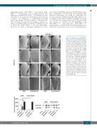
Figure 5. Dasatinib rescues BCR-ABL1p210 driven eye defect and shows target speci- ficity in vivo. Scanning electron micrographs of adult Drosophila compound eyes from flies fed on 0.03% DMSO only or dasatinib. Posterior is to the left. GMR-GAL4>w1118 were used as control. E-H and M-P are high mag- nification of the posterior end of the eye in A-D and I-L respectively (692x). Normal development in control flies fed on DMSO (A,E, I, M) or dasatinib is observed. BCR- ABL1p210 (C, G) and BCR-ABL1p210/T315I (K, O) expressing flies fed on DMSO show charac- teristic defective area with loss of ommatidi- al facets. Area is marked with a representa- tive dashed line. Ommatidial development in this area was restored with BCR-ABL1p210 flies fed on 20 μM dasatinib (D, H). Compare to (C, G). BCR-ABLp210/T315I flies showed no restoration of ommatidial devel- opment (L, P). Compare to (K, O). Lower left panel represents measurement of the pos- terior eye defect area (μm2). Data repre- sents mean ± SEM. ****, P<0.0001. Lower right panel is a representative Western blot of the expression of BCR-ABL1 and phos- phorylated levels in transgenic untreated and treated adult fly heads. Genotypes indi- cated are under the control of eye specific promoter GMR-GAL4.
haematologica | 2020; 105(2)
393
