
Page 148 - 2020_02-Haematologica-web
P. 148
V. Caraffini et al.
data indicate that RKIP loss enhances RAS-MAPK/ERK signaling on the one hand and increases GM-CSF-induced myelomonocytic lineage commitment and differentiation of HSPC on the other. Furthermore, they suggest that RKIP exerts its role in terminal myelomonocytic differen- tiation by acting as a rheostat that modulates the sensitiv- ity to external stimuli, such as 1,25D3 and growth factors, respectively.
Rkip deletion aggravates myeloproliferation and the development of a myelomonocytic myeloproliferative disease in Ras-mutated mice
Increased myelomonocytic lineage commitment is a key step in myeloid leukemogenesis. However, Rkip-/- mice failed to develop myeloid neoplasias in our study (data not shown). As the functional assays delineating the role of RKIP in myelomonocytic differentiation suggested that RKIP rather acts as an amplifier of activated GM-CSF/RAS signaling, we next crossed Rkip-/- mice with Mx1-Cre/Nras-
mutated animals (Online Supplementary Figure S5). The Mx1-Cre/Nras was chosen because: (i) RKIP loss and RAS-signaling mutations have previously been shown to co-occur in AML;12,20,22 and (ii) RKIP loss has been demon- strated to potentiate the oncogenic effects of RAS-signal- ing mutations in functional in vitro assays.12,20 Interestingly, Mx1-Cre/Nras mice on a pure C57BL/6 background devel- op a myeloproliferation that preferentially affects the myelomonocytic lineages. However, previously published data have demonstrated that these mice ultimately suc- cumb to histiocytic sarcomas (HS) and only randomly develop a full blown MPD.8 In the current study, we elec- tively analyzed mice at an age of six months after the first pIpC injection and thereby observed that myeloprolifera- tion was aggravated in Mx1-Cre/Nras/Rkip-/- mice, who consistently demonstrated splenomegaly, as well as increased myeloid infiltration of bone marrow, spleen and liver as compared to Nras/Rkip+/+ and to Mx1- Cre/Nras/Rkip+/+ animals (Figures 4A and B, and 5). In addi-
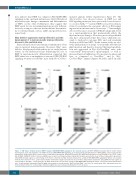
AB
CD
Figure 6. RAF kinase inhibitor protein (RKIP) regulates RAS-MAPK/ERK signaling in the myeloid system. (A) RKIP shRNA knockdown (KD) in CD34+ human hematopoietic stem and progenitor cells (HSPC) increased RAS-MAPK/ERK signaling, as measured by the phosphorylation of ERK (pERK). (Left) A representative immunoblot. Graph represents the mean of three independent experiments ±Standard Deviation (SD); pERK intensity is given as x-fold change to the CD34+ control KD. (B) RKIP shRNA KD in HL-60 increased pERK levels as well. (Left) Representative immunoblot. Graph represents the mean of three independent experiments ±SD; pERK intensity is given as x-fold change to the HL-60 control KD. (C) Rkip deletion enhanced the activity of RAS-MAPK/ERK signaling in CD11b+ cells isolated from the bone marrow of mice. (Left) A representative immunoblot. Graph represents the mean of three independent experiments ±SD; pERK intensity is given as x-fold change to the Rkip+/+ control genotype. (D) Rkip deletion also increased the activity of RAS-MAPK/ERK signaling in CD11b+ cells isolated from Nras-mutated mice. A representative immunoblot and the graph is presented as described above, Mx1-Cre/Nras/Rkip+/+ were used as control group. Mice experiments were per- formed using n=3 mice for each genotype. Statistical significance was evaluated by Student’s t-test in all cases.
382
haematologica | 2020; 105(2)
