
Page 283 - Haematologica Atlas of Hematologic Cytology
P. 283
CHAPtER 35 - Thrombotic microangiopathies
Chapter 35. THROMBOTIC MICROANGIOPATHIES
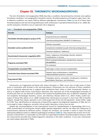
The term thrombotic microangiopathy (TMA) describes a condition characterized by arteriolar and capillary thrombosis resulting in microangiopathic hemolytic anemia, thrombocytopenia and frequent organ injury. Qui- te different conditions can induce TMA by different pathogenetic mechanisms (Table 1), but all of them share thrombocytopenia and red cell anisopoikilocytosis with schistocytes in peripheral blood (Scully et al., 2018). Blo- od film evaluation, therefore, has an important role in diagnosis.
Table 1. Thrombotic microangiopathies (TMA).
Disorder
Subtypes
Thrombo c thrombocytopenic purpura (TTP)
Acquired (immune-mediated)
Inherited predisposi on (ADAMTS-13 de ciency)
Hemoly c uremic syndrome (H S)
Infec on-associated
Complement-mediated (usually inherited predisposi on)
Inherited cobalamin C de ciency
Disseminated intravascular coagula on (DI )
Sepsis, malignancy, hematologic disorder, obstetric complica ons, or trauma
Pregnancy associated TM
HELLP syndrome (hemolysis, elevated liver enzymes, low platelets)
Pre-eclampsia
Transplanta on associated TM
Solid organ or bone marrow transplanta on
onnec ve ssue disease associated TM
Systemic lupus erythematosus, scleroderma, an phospholipid syndrome, vasculi s
Drug induced TM
Ticlopidine, quinine, simvasta n, trimethoprim, interferon, gemcitabine, bleomycin, and mitomycin
Schistocytes are fragmented red blood cells whose formation in TMAs occurs due to high levels of shear stress in association with thrombi in the microvasculature. Schistocytes are not exclusive to these conditions but are commonly observed also in subjects with mechanical heart valves or other intravascular devices, as well as in subjects with chronic renal disease. Moreover, they represent one of the morphological anomalies of erythrocytes that can be observed in patients with marked anisopoikilocytosis due to different neoplastic and non-neoplastic hematologic disorders, cancer, and infectious diseases (Huh et al., 2013). Finally, careful eva- luation of blood films (Burns et al., 2004) revealed that schistocytes are present in more than 50% of healthy subjects, though they always represent a very small proportion of the red cell population (mean 0.05%, range 0-0.27%). Thus, identification of schistocytes on blood films supports the diagnostic hypothesis of TMA, but it is not enough to make a diagnosis, and other clinical and/or laboratory criteria must be met.
Despite the recommendations of the International Council for Standardization in Haematology (Zini et al., 2012), quantification of schistocytes remains highly operator-dependent, and this can affect the comparison of measurements obtained by different authors. Despite this limitation, we consider it useful to report the data obtained by different authors in different pathological conditions (Table 2). They indicate that at least 1% of schi- stocytes in blood films is a reasonable cut-off value for suspecting thrombotic thrombocytopenic purpura, while this measurement provides few reliable diagnostic indications in the other conditions. In any case, schistocytes are nearly always observed in TMAs.
270
