
Page 282 - Haematologica Atlas of Hematologic Cytology
P. 282
AB
CD
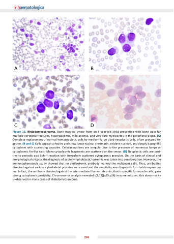
Figure 13. Rhabdomyosarcoma. Bone marrow smear from an 8-year-old child presenting with bone pain for multiple vertebral fractures, hypercalcemia, mild anemia, and very rare myelocytes in the peripheral blood. (A) Complete replacement of normal hematopoietic cells by medium-large sized neoplastic cells, often grouped to- gether. (B and C) Cells appear cohesive and show loose nuclear chromatin, evident nucleoli, and deeply basophilic cytoplasm with coalescing vacuoles. Cellular outlines are irregular due to the presence of numerous lumps or cytoplasmic fin-like tails. Many cytoplasmic fragments are scattered on the smear. (D) Neoplastic cells are posi- tive to periodic acid Schiff reaction with irregularly scattered cytoplasmic granules. On the basis of clinical and morphological criteria, the diagnosis of acute lymphoblastic leukemia was taken into consideration. However, the immunophenotypic study showed that no antileukemic antibody marked the malignant cells. Thus, antibodies directed against various cytoskeletal proteins were used and the reactivity was diagnostic for rhabdomyosarco- ma. In fact, the antibody directed against the intermediate filament desmin, that is specific for muscle cells, gave strong cytoplasmic positivity. Chromosomal analysis revealed t(2;13)(q35;q14) in some mitoses; this abnormality is observed in many cases of rhabdomyosarcoma.
269
