
Page 61 - 2019_12-Haematologica-web
P. 61
KLF1 mutation in human anemia
this phenotypic effect, even though both alleles are expressed (Online Supplementary Figure S1B), total KLF1 expression is lower in the CDA erythroid cell (Figure 2B) verified by direct RT-qPCR (data not shown).
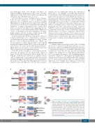
Cell cycle effects are less straightforward to interpret. Both cell cycle stimulators as well as inhibitors play important roles at different stages of erythroid terminal differentiation.18 In the present case (Figure 2C), E2F2 and E2F4 levels are overall lower in the patient cells, while p21 (CDKN1A) is higher. Surprisingly in this context, TP53 levels are lower in the patient cells, suggesting that the increase in p21 occurs independently of p53.44 Expression of pro- or anti-apoptosis protein-coding genes are mini- mally changed, with the exception of BCL2 and possibly PIM1, whose levels are higher in the CDA cell.
Given the phenotypic problems with nuclear extrusion, bi-nuclei, and bridges, it is of interest that a number of cytokinesis/mitosis proteins implicated in these cellular processes such as pleckstrin (PLEK2) and TRIM58 are lower in expression (Figure 2D). In a link to CDA type III patients that express mutant KIF23, levels of this gene are decreased (as also noted in the KLF1 hydrops patient).29 Other CDA-associated genes such as CDAN1 (type I) and SEC23B (type II) are not significantly altered.
Given the extent of disruption of iron utilization in the CDA type IV patients, we also queried this subset of genes as well, and found varied effects (Figure 2E). For example, erythroferrone (FAM132B), a regulator of hep- cidin, is dramatically down-regulated in the CDA patient (and thus different from CDA type II45), as are the transfer- rin receptors (TFR2 and TFRC). Although levels of the iron exporter ferroportin (SCL40A1) and the heme regulator
AB
SLC48A1 are not significantly altered, this combination likely leads to a net inability to mobilize and incorporate iron and heme in the CDA patient, particularly when cou- pled to the low expression of the ABCB6 gene (noted ear- lier) that would decrease activity of this important por- phyrin importer. These conditions could account for the anisopoikilocytosis seen in spite of normal MCV. Ferritin levels (FTL and FTH1) are increased, which may con- tribute to the high levels of stored iron observed in the patient. By the same token, increased heme oxygenase (HMOX1) could account for the hyperbilirubinemia seen in all patients with type IV CDA. Alternatively, increased bilirubin may be secondary to hemolysis, with HMOX1 expression increased in response. These effects are remi- niscent of those resulting from the mouse Nan mutation, which contains a change in the same amino acid (albeit to D) and leads to extensive iron and heme regulation disrup- tion.33
Differentiation deficits
To identify expression changes in the CDA cell during the process of proliferation and differentiation, we com- pared genes expressed ≥5 FPKM (Fragments Per Kilobase of transcript per Million) in all samples. Individual Venn comparisons reveal that the d11 and d15 proliferating samples each overlap >72%, but the differentiation d5 set does not continue this pattern, only overlapping approxi- mately 55% (Figure 3A). This suggests that terminal differ- entiation does not proceed normally in the CDA erythroid cell. To more directly address this, we analyzed RNA expression of our samples and compared it to that from cells undergoing normal human erythropoiesis.39,46,47 At
C
D
E
Figure 2. Relative expression levels following RNA-seq analysis.
Fragments Per Kilobase of transcript per Million (FPKM) expression val- ues were obtained following erythroid proliferation [day d11 and d15] and differentiation (d5) of normal (wild-type, WT) or patient (CDA) periph- eral blood mononuclear cells (Online Supplementary Table S1). The pro- liferation/differentiation series is grouped together and color-coded based on expression of single samples taken at each time point. Color- coding was based on relative expression of samples within each group. Genes were selected based on their importance in: (A) cell surface expression, structural or membrane integrity; (B) β-like globin gene regu- lation; (C) cell cycle and apoptosis; (D) cytokinesis and mitosis; and (E) iron utilization and storage.
haematologica | 2019; 104(12)
2375
