
Page 60 - 2019_12-Haematologica-web
P. 60
L. Varricchio et al.
Results
Establishment of expansion protocol
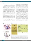
Our patient had been analyzed with respect to hemato- logic parameters such as red cell surface expression, peripheral blood smear, and globin expression pattern.5 For the present study, erythroid cells were expanded from PBMC using an ex vivo culture system. This culture system contains stimulatory cytokines along with the critical inclusion of dexamethasone to enable efficient expansion of the small number of erythroid progenitors present in a typical mononuclear cell preparation.36,37 Using this proto- col we established and expanded erythroid cells from the CDA type IV patient in parallel with a normal control. Morphological examination (Figure 1) reveals that the expanded patient cells exhibit bi- and multi-nucleated cells with abnormal nuclei (approx. 40%) as seen in the original bone marrow and blood smears of the patient.5 These are observed in approximately 5% of cells from the normal control.
Similar to the limitation in studying erythropoiesis in murine Klf1-null cells,34 expression of many of the cell sur- face markers for differentiation are quite low and not informative for staging purposes, as they are KLF1 targets. As a result, we assessed and compared the range of cellu- lar morphologies in the d11 proliferating samples, and find these are not significantly different (Figure 1C).
Global dysregulation of erythroid genes in the CDA cell
Given the availability of cells from only a single patient, we aimed to analyze cells at three time points to increase
AB
the robustness of our data. As a result, the PBMC under- went a 2-step culture: one for proliferation, with harvests at d11 and d15; the second for differentiation started at d11 and harvested after an additional five days. RNA was isolated from all samples and analyzed for gene expres- sion via deep RNA sequencing. Focusing our analysis on selected targets, we find a radical alteration of gene expression that covers cell cycle, membrane protein, and globin switching deficits, all congruent with the pheno- typic properties of CDA type IV cells (Figure 2). For exam- ple, although expression of some cell surface molecules (CD47, CD55, CD58) are minimally affected as noted before by FACS analysis of primary patient cells4 (and thus serve as controls), GYPA (CD235a) expression is dramati- cally lower. This is mimicked by radically low levels of KLF1-regulated structural proteins such as ICAM4, tropo- modulin (TMOD1), and band 4.2 (EPB42), likely con- tributing to the fragility of patients’ red cells and their membrane abnormalities. Transport proteins such as anion transporter band 3 (SLC4A1), ion channel PIEZO1, potassium-calcium channel KCNN4, ABC transporter ABCB6, and aquaporin water channel AQP3 exhibit extremely low levels of expression (Figure 2A).
C
Cells from the CDA patient express high levels of g-glo- bin, reaching approximately 90% of total globin (Online Supplementary Figure S1A). With respect to regulation of globin switching, two KLF1 targets that act as g-globin repressors were checked.41-43 BCL11A levels are decreased but not that of ZRF/Pokemon (ZBTB7A) (Figure 2B), sug- gesting that the increase in g-globin follows derepression as a result of the drop in BCL11A protein. Contributing to
Figure 1. Morphological assessment. (A) Bone marrow aspirate of patient. (B) (Left) Cytospin of patient periph- eral blood mononuclear cells after expansion for 11 days; (right) similar analysis for normal patient sample per- formed in parallel. Analysis of multiple slides revealed abnormal nuclei in 42.7% of CDA cells, and 5.6% of nor- mal cells. (C) Quantification via cellular morphology of wild-type (WT) and patient cells categorized as proery- throblast (ProE), basophilic erythroblast (Baso), polychromatophillic erythroblast (Poly), or orthchromatic ery- throblast (Ortho). Multiple cytospin slides from two experiments were separately quantified by two investigators.
2374
haematologica | 2019; 104(12)
